This paper reports the analysis of the intramolecular OH stretching band obtained by Fourier Transform Infrared (FTIR) spectroscopy measurements. In order to characterize the effect of montmorillonite on the properties of Bovine Serum Albumin (BSA) the two-state model is adopted for the analysis of the OH stretching band. We assume that the OH stretching can be divided into two different states of inter-molecular bonding. The results of this experimental work confirm that the montmorillonite leads to a stabilization of the BSA structure. Also, the analysis of the spectra temperature dependence shows a montmorillonite-induced higher thermal stability of the BSA in respect to pristine BSA. Thus, this paper highlights the effectiveness of montmorillonite as thermal bio-protector. A FTIR analysis was carried out to investigate the interaction of Montmorillonite with BSA. Two different approaches, i.e. Spectral Distance and Wavelet analyses, constitute two effective and innovative approaches for the characterization of the thermal properties of pristine BSA and of BSA in the presence of Montmorillonite. The results allowed us to consider as BSA in the presence of Montmorillonite has a lower spectral sensitivity when the temperature changes and, therefore, the role of Montmorillonite as a thermal bio-protector is motivated.
Citation: Maria Teresa Caccamo, Giuseppe Mavilia, Letterio Mavilia, Pietro Calandra, Domenico Lombardo, Salvatore Magazù. Thermal investigation of montmorillonite/BSA by fourier transform infrared spectroscopy measurements
This paper reports the analysis of the intramolecular OH stretching band obtained by Fourier Transform Infrared (FTIR) spectroscopy measurements. In order to characterize the effect of montmorillonite on the properties of Bovine Serum Albumin (BSA) the two-state model is adopted for the analysis of the OH stretching band. We assume that the OH stretching can be divided into two different states of inter-molecular bonding. The results of this experimental work confirm that the montmorillonite leads to a stabilization of the BSA structure. Also, the analysis of the spectra temperature dependence shows a montmorillonite-induced higher thermal stability of the BSA in respect to pristine BSA. Thus, this paper highlights the effectiveness of montmorillonite as thermal bio-protector. A FTIR analysis was carried out to investigate the interaction of Montmorillonite with BSA. Two different approaches, i.e. Spectral Distance and Wavelet analyses, constitute two effective and innovative approaches for the characterization of the thermal properties of pristine BSA and of BSA in the presence of Montmorillonite. The results allowed us to consider as BSA in the presence of Montmorillonite has a lower spectral sensitivity when the temperature changes and, therefore, the role of Montmorillonite as a thermal bio-protector is motivated.

| [1] | Leach S (2015) Origins of life. Adv Chem Phys 157: 293-313. |
| [2] | Haldane JBS (1929) The origin of life. Rationalist Annual 148: 3-10. |
| [3] |
Bernal JD (1949) The physical basis of life. P Phys Soc 62: 597. 
|
| [4] | Cains-Smith AG (1982) Genetic Takeover. Cambridge: Cambridge University Press. |
| [5] |
Lehn JM (2002) Toward complex matter: Supramolecular chemistry and self-organization. PNAS 99: 4763-4768.
|
| [6] |
Lehn JM (2002) Toward self-organization and complex matter. Science 295: 2400-2403.
|
| [7] |
Bada JL, Lazcano A (2003) Perceptions of science. Prebiotic soup-revisiting the Miller experiment. Science 300: 745-746.
|
| [8] |
Watson JD, Crick FHC (1953) Molecular structure of nucleic acids. Nature 171: 737-738.
|
| [9] |
Troland LT (1914) The chemical origin and regulation of life. The Monist 22: 92-133.
|
| [10] |
Muller HJ (1961) Genetic nucleic acid: Key material in the origin of life. Persp Biol Med 5: 1-23.
|
| [11] |
Gilbert W (1986) Origin of life: The RNA world. Nature 319: 618-618.
|
| [12] |
Chaabani H (2015) Man creation had began since the creation of the first biological material very likely in Clay. Int J Mod Anthrop 8: 49-65.
|
| [13] |
Luisi PL (2006) The Emergence of Life. From Chemical Origins to Synthetic Biology. Cambridge: Cambridge University Press.
|
| [14] |
Szostak JW, Bartel DP, Luisi PL (2001) Synthesizing life. Nature 409: 387-390.
|
| [15] |
Nielsen PE, Egholm M, Berg RH, et al. (1991) Sequence-selective recognition of DNA by strand displacement with a thymine-substituted polyamide. Science 254: 1497-1500.
|
| [16] | Lazcano A (2010) Historical development of origins research. Cold Spring Harbor Persp BioL 2: a002089. |
| [17] |
Wachtershauser G (1988) Before enzymes and templates, a theory of surface metabolism. Microbiol Rev 52: 452-484.
|
| [18] | Haldane JBS (1954) The origins of life. New Biol 16: 12-27. |
| [19] |
Muller HJ (1966) The gene material as the initiator and the organizing basis of life. Am Naturalist 100: 493-517.
|
| [20] | Raven PH, Johnson GB, Mason KA, et al. (2014) The nature of molecules and properties of water. Biology . New York: McGraw-Hill 17-30. |
| [21] | Reece JB, Urry LA, Cain ML, et al. (2011) Water and life. Campbell Biology . San Francisco: Pearson 44-54. |
| [22] |
Caetano-Anollés G, Wang M, Caetano-Anollés D (2013) Structural phylogenomics retrodicts the origin of the genetic code and uncovers the evolutionary impact of protein flexibility. PLoS One 8: e72225.
|
| [23] |
Eigen M, Lindemann BF, Tietze M, et al. (1989) How old is the genetic code? Statistical geometry of tRNA provides an answer. Science 244: 673-679.
|
| [24] |
Ertem G, Ferris JP (2000) Sequence and regio-selectivity in the montmorillonite catalyzed synthesis of RNA. Orig Life Evo Biosph 30: 411-422.
|
| [25] | Farias ST, do Rego TG, José MV (2014) Evolution of transfer RNA and the origin of the translation system. Fron Genet 5: 303-306. |
| [26] |
Ghadiri M, Chrzanowski W, Rohanizadeh R (2015) Biomedical applications of cationic clay minerals. RSC Adv 5: 29467-29481.
|
| [27] |
de Paiva LB, Morales AR, Díaz FRV (2008) Organoclays: properties, preparation and applications. App Clay Sci 42: 8-24.
|
| [28] | Datta M (2013) Clay–polymer nanocomposites as a novel drug carrier: Synthesis, characterization and controlled release study of Propranolol Hydrochloride. Appl Clay Sci 80–81: 85-92. |
| [29] |
Xiang Y, Villemure G (1996) Electrodes modified with synthetic clay minerals: electrochemistry of cobalt smectites. Clays Clay Miner 44: 515-521.
|
| [30] |
Patel HA, Somani RS, Bajaj HC, et al. (2006) Nanoclays for polymer nanocomposites, paints, inks, greases and cosmetics formulations, drug delivery vehicle and waste water treatment. Bull Mater Sci 29: 133-145.
|
| [31] |
Yu WH, Li N, Tong DS, et al. (2013) Adsorption of proteins and nucleic acids on clay minerals and their interactions: A review. Appl Clay Sci 80–81: 443-452.
|
| [32] | Stănescu VN, Olteanu M, Florea-Spiroiu M, et al. (2008) Fractal properties of collagen/chitosan/montmorillonite membranes. Rev Roum Chim 54: 767-771. |
| [33] | Raussell-Colom JA (1987) Reactions of clays with organic substances. Chem Clays Clay Miner, Mineral Soc : 412-415. |
| [34] |
Bergaya F, Lagaly G (2001) Surface modifications of clay minerals. Appl Clay Sci 19: 1-3.
|
| [35] |
Caccamo MT, Mavilia G, Mavilia L, et al. (2020) Self-assembly processes in hydrated montmorillonite by FTIR investigations. Materials 13: 1100.
|
| [36] | Maina E, Wanyika H, Gachanja A (2016) Natural pyrethrum extracts photo-stabilized with organo clays. J Sci Res Rep 9: 1-20. |
| [37] | Ismadji S, Soetaredjo F, Ayucitra A, et al. (2015) Natural clay minerals as environmental cleaning agents. J Clay Mat Environ Rem 8: 5-37. |
| [38] |
Celis R, HermosÍN C, Cornejo L, et al. (2010) Clay–herbicide complexes to retard picrolam leaching in soil. Int J Environ Analyt Chem 82: 503-517.
|
| [39] |
Xi Y, Frost RL, He H (2007) Modification of the surfaces of Wyoming montmorillonite by the cationic surfactants alkyl trimethyl, dialkyl dimethyl and trialkylmethyl ammonium bromide. J Coll Interf Sci 305: 150-158.
|
| [40] |
Savelyev YV, Gonchar AN (2019) Exfoliation of montmorillonite in polymer matrix and its influence on the nanocomposites properties. Polym J 41: 149-158.
|
| [41] |
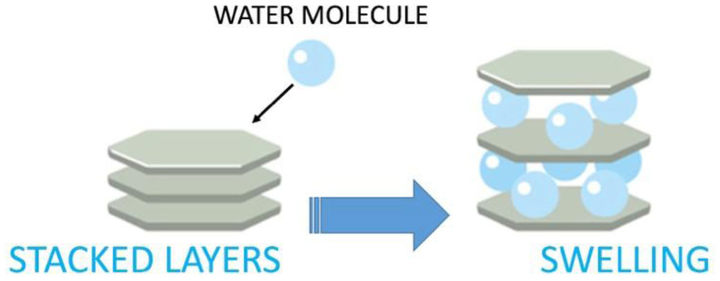
Emiel JM, Hensen SB (2002) Why clay swell. J Phys Chem B 106: 12664-12667.
|
| [42] |
Tyagi B, Chudasama CD, Jasra RV (2006) Determination of structural modification in acid activated montmorillonite clay by FT-IR spectroscopy. Spectrochim Acta A 64: 273-278.
|
| [43] |
Temuujin J, Jadambaa T, Burmaa G, et al. (2004) Characterisation of acid activated montmorillonite clay from tuulant (Mongolia). Ceram Int 30: 251-255.
|
| [44] |
Bonaccorsi L, Calandra P, Kiselev MA, et al. (2013) Self-assembly in poly(dimethylsiloxane)-poly(ethylene oxide) block copolymer template directed synthesis of linde type A zeolite. Langmuir 29: 7079-7086.
|
| [45] |
Bonaccorsi L, Calandra P, Amenitsch H, et al. (2013) Growth of fractal aggregates during template directed SAPO-34 zeolite formation. Microporous Mesoporous Mat 167: 3-9.
|
| [46] |
Bonaccorsi L, Lombardo D, Longo A, et al. (2009) Dendrimer template directed self-assembly during zeolite formation. Macromol 42: 1239-1243.
|
| [47] |
Chen C, Wylie RAL, Klinger D, et al. (2017) Shape control of soft nanoparticles and their assemblies. Chem Mater 29: 1918-1945.
|
| [48] |
Kiselev MA, Lombardo D (2017) Structural characterization in mixed lipid membrane systems by neutron and X-ray scattering. Biochem Biophys Acta-Gen Sub 1861: 3700-3717.
|
| [49] |
Lombardo D, Munaò G, Calandra P, et al. (2019) Evidence of pre-micellar aggregates in aqueous solution of amphiphilic PDMS–PEO block copolymer. PCCP 21: 11983-11991.
|
| [50] |
Rao Y, Blanton TN (2008) Polymer nanocomposites with a low thermal expansion coefficient. Macromolecules 41: 935-941.
|
| [51] |
Huang W, Ferris JP (2006) One-step, regioselective synthesis of up to 50-mers of RNA oligomers by montmorillonite catalysis. J Am Chem Soc 128: 8914-8919.
|
| [52] |
He H, Guo J, Xie X, et al. (2002) Microstructural study of acid-activated montmorillonite from Choushan, China. Clay Miner 37: 337-344.
|
| [53] |
Ferris JP (2005) Mineral calalysis and prebiotic synthesis: Montmorillonite-catalyzed formation of RNA. Elements 1: 145-149.
|
| [54] |
Joshi PC, Pitsch S, Ferris JP (2000) Homochiral selection in the montmorillonite-catalyzed and uncatalyzed prebiotic synthesis of RNA. Chem Comm : 2497-2498.
|
| [55] |
Mazo MA, Manevitch LI, Gusarova EB, et al. (2008) Molecular dynamics simulation of thermomechanical properties of montmorillonite crystal. 3. Montmorillonite crystals with PEO oligomer intercalates. J Phys Chem B 112: 3597-3604.
|
| [56] |
Adams JM (1987) Synthetic organic chemistry using pillared, cation-exchanged and acid-treated montmorillonite catalysts—A review. Appl Clay Sci 1987: 309-342.
|
| [57] |
Miyakawa S, Ferris JP (2003) Sequence-and regioselectivity in the montmorillonite-catalyzed synthesis of RNA. J Am Chem Soc 125: 8202-8208.
|
| [58] |
Kawamura K, Ferris JP (1999) Clay catalysis of oligonucleotide formation: kinetics of the reaction of the 5′-phosphorimidazolides of nucleotides with the non-basic heterocycles uracil and hypoxanthine. Orig Life Evol Biosph 29: 563-591.
|
| [59] |
Joshi PC, Aldersley MF, Delano JW, et al. (2009) Mechanism of montmorillonite catalysis in the formation of RNA oligomers. J Am Chem Soc 131: 13369-13374.
|
| [60] |
Knauth LP (1998) Salinity history of the earth's early ocean. Nature 395: 554-555.
|
| [61] |
Knauth LP (2005) Temperature and salinity history of the precambrian ocean: Implications for the course of microbial evolution. Palaegeogr Palaeoclimatol Palacoecol 2190: 53-69.
|
| [62] |
Hren MT, Tice MM, Chamberlain CP (2009) Oxygen and hydrogen isotope evidence for a temperature 3.42 billion years ago. Nature 462: 205-208.
|
| [63] |
Zhou ZJ, Cameron S, Kadatz B, et al. (1997) Clay swelling diagrams: their applications in formation damage control. SPE J 2: 99-106.
|
| [64] | Sposito G (1984) The Surface Chemistry of Soils. New York: Oxford University Press 115-2208. |
| [65] |
Joshi PC, Aldersley MF, Delano JW, et al. (2009) Mechanism of montmorillonite catalysis in the formation of RNA oligomers. J Am Chem Soc 131: 13369-13374.
|
| [66] |
Hanczyc MM, Fujikawa SM, Szostak JW (2003) Experimental models of primitive cellular compartments: Encapsulation, growth, and division. Science 302: 618-622.
|
| [67] |
Hanczyc MM, Mansy SS, Szostak JW (2007) Mineral surface directed membrane assembly. Orig Life Evol Biosph 37: 67-82.
|
| [68] |
Mansy SS, Szostak JW (2008) Thermostability of model protocell membranes. Proc Natl Acad Sci USA 105: 3351-13355.
|
| [69] | Guerrini L, Alvarez-Puebla RA, Pazos-Perez N (2018) Surface modifications of nanoparticles for stability in biological fluids. Materials 1154. |
| [70] |
Lombardo D (2009) Liquid-like ordering of negatively charged poly (amidoamine) (PAMAM) dendrimers in solution. Langmuir 25: 3271-3275.
|
| [71] |
Lombardo D (2014) Modeling dendrimers charge interaction in solution: Relevance in biosystems. Biochem Res Int 2014: 837651.
|
| [72] |
Lombardo D, Calandra P, Magazù S, et al. (2018) Soft nanoparticles charge expression within lipid membranes: The case of amino terminated dendrimers in bilayers vesicles. Colloids Surf, B 170: 609-616.
|
| [73] |
Hrenovic J, Milenkovic J, Ivankovic T, et al. (2012) Antibacterial activity of heavy metal-loaded natural zeolite. J Hazard Mater 201: 260-264.
|
| [74] |
Hu CH, Xu ZR, Xia MS (2005) Antibacterial effect of Cu2+-exchanged montmorillonite on Aeromonas hydrophila and discussion on its mechanism. Vet Microbiol 109: 83-88.
|
| [75] |
Magana SM, Quintana P, Aguilar DH, et al. (2008) Antibacterial activity of montmorillonites modified with silver. J Mol Catal A Chem 281: 192-199.
|
| [76] |
Malachová K, Praus P, Rybková Z, et al. (2011) Antibacterial and antifungal activities of silver, copper and zinc montmorillonites. Appl Clay Sci 53: 642-645.
|
| [77] |
Morrison KD, Underwood JC, Metge DW, et al. (2014) Mineralogical variables that control the antibacterial effectiveness of a natural clay deposit. Environ Geochem Health 36: 613-631.
|
| [78] |
Tong G, Yulong M, Peng G, et al. (2005) Antibacterial effects of the Cu (II)-exchanged montmorillonite on Escherichia coli K88 and Salmonella choleraesuis. Vet Microbiol 105: 113-122.
|
| [79] |
Williams LB, Haydel SE (2010) Evaluation of the medicinal use of clay minerals as antibacterial agents. Int Geol Rev 52: 745-770.
|
| [80] |
Liyanapatirana C, Gwaltney SR, Xia K (2009) Transformation of triclosan by Fe (III)-saturated montmorillonite. Environ Sci Technol 44: 668-674.
|
| [81] |
Qin C, Troya D, Shang C, et al. (2014) Surface catalyzed oxidative oligomerization of 17 β-estradiol by Fe3+-saturated montmorillonite. Environ Sci Technol 49: 956-964.
|
| [82] |
Qin C, Chen C, Shang C, et al. (2018) Fe3+-saturated montmorillonite effectively deactivates bacteria in wastewater. Sci Total Environ 622–623: 88-95.
|
| [83] |
Majorek KA, Porebski PJ, Dayal A, et al. (2012) Structural and immunologic characterization of bovine, horse, and rabbit serum albumins. Mol Imm 52: 174-182.
|
| [84] |
Benkő M, Varga N, Sebők D, et al. (2015) Bovine serum albumin-sodium alkyl sulfates bioconjugates as drug delivery systems. Coll Surf B Biointerf 130: 126-132.
|
| [85] |
Xu W, Peng J, Ni D, et al. (2020) Preparation, characterization and application of levan/montmorillonite biocomposite and levan/BSA nanoparticle. Carb Polym 234: 115921.
|
| [86] |
Minutoli L, Altavilla D, Bitto A, et al. (2008) Trehalose: A biophysics approach to modulate the inflammatory response during endotoxic shock. Eur J Pharm 589: 272-280.
|
| [87] |
Maisano G, Majolino D, Migliardo P, et al. (1993) Sound velocity and hydration phenomena in aqueous polymeric solutions. Mol Phys 78: 421-435.
|
| [88] |
Magazù S, Migliardo F, Affouard F, et al. (2010) Study of the relaxational and vibrational dynamics of bioprotectant glass-forming mixtures by neutron scattering and molecular dynamics simulation. J Chem Phys 132: 184512.
|
| [89] |
Adamo A, Calabrò E, Magazù S (2019) Thermostabilization of BSA in TMAO water mixtures by infrared spectroscopy. Curr Chem Biol 13: 49-59.
|
| [90] | Magazù S, Caccamo MT (2019) Study of trehalose effects on bsa solutions. Trehalose: Sources, Chemistry and Applications . Hauppauge: Nova Science Publishers 43-62. |
| [91] |
Magazù S, Migliardo F, Benedetto A, et al. (2013) Protein dynamics by neutron scattering: The protein dynamical transition and the fragile-to-strong dynamical crossover in hydrated lysozyme. Chem Phys 424: 26-31.
|
| [92] |
Magazù S, Migliardo F, Benedetto A (2011) Puzzle of protein dynamical transition. J Phys Chem B 115: 7736-7743.
|
| [93] |
Fenimore PW, Frauenfelder H, Magazù S, et al. (2013) Concepts and problems in protein dynamics. Chem Phys 424: 2-6.
|
| [94] |
Magazù S, Calabro E, T Caccamo M, et al. (2016) The shielding action of disaccharides for typical proteins in aqueous solution against static, 50 Hz and 1800 MHz frequencies electromagnetic fields. Curr Chem Biol 10: 57-64.
|
| [95] | Magazù S, Migliardo F, Benedetto A, et al. (2013) Bioprotective effects of sucrose and trehalose on proteins. Sucrose: Properties, Biosynthesis and Health Implications . Hauppauge: Nova Science Publishers 43-62. |
| [96] | Cannuli A, Caccamo MT, Magazù S (2018) Modeling and self-organization dynamics of aggregation processes in acoustically levitated disaccharides solutions. AAPP Atti della Accademia Peloritana dei Pericolanti, Classe di Scienze Fisiche, Matematiche e Naturali 96: 3. |
| [97] |
Squire PG, Maser P, O'Konski CT (1968) The hydrodynamic properties of bovine serum albumin monomer and dimmer. Biochem 7: 4261-4272.
|
| [98] |
De Paz RA, Dale DA, Barnett CC, et al. (2002) Effects of drying methods and additives on the structure, function, and storage stability of subtilisin: role of protein conformation and molecular mobility. Enzyme Microb Technol 31: 765-774.
|
| [99] |
Xu Y, Hanna MA (2008) Morphological and structural properties of two-phase coaxial jet electrosprayed BSA-PLA capsules. J Microencapsul 25: 469-477.
|
| [100] |
Kopac T, Bozgeyik K, Yener J (2008) Effect of pH and temperature on the adsorption of bovine serum albumin onto titanium dioxide. Coll Surf Physicochem Engin Aspects 322: 19-28.
|
| [101] |
Yohannes G, Wiedmer SK, Elomaa M, et al. (2010) Thermal aggregation of bovine serum albumin studied by asymmetrical flow field-flow fractionation. Anal Chim Acta 675: 191-198.
|
| [102] |
Alkan M, Demirbas O, Dogan M, et al. (2006) Surface properties of bovine serum albumin—adsorbed oxides: Adsorption, adsorption kinetics and electrokinetic properties. Micro Meso Materials 96: 331-340.
|
| [103] |
Magazù S, Migliardo F, Benedetto A (2011) Elastic incoherent neutron scattering operating by varying instrumental energy resolution: Principle, simulations, and experiments of the resolution elastic neutron scattering (RENS). Rev Sci Instr 82: 105115.
|
| [104] |
Migliardo F, Magazù S, Caccamo MT (2013) Infrared, Raman and INS studies of poly-ethylene oxide oligomers. J Mol Struc 1048: 261-266.
|
| [105] |
Calabrò E, Condello S, Currò M, et al. (2013) Effects of low intensity static magnetic field on FTIR spectra and ROS production in SH-SY5Y neuronal-like cells. Bioelectromagn 34: 618-629.
|
| [106] |
Varga B, Migliardo F, Takacs E, et al. (2008) Neutron scattering studies on dUTPase complex in the presence of bioprotectant systems. Chem Phys 345: 250-258.
|
| [107] |
Cannuli A, Caccamo MT, Castorina G, et al. (2018) Laser techniques on acoustically levitated droplets. EPJ Web of Confer 167: 5010.
|
| [108] | Egli M (2016) Diffraction techniques in structural biology. Curr Protoc Nucleic Acid Chem 65: 7.13.1-7.13.41. |
| [109] |
Dong M, Husale S, Sahin O (2009) Determination of protein structural flexibility by microsecond force spectroscopy. Nature nanotechn 4: 514-517.
|
| [110] |
Becker W, Bhattiprolu KC, Gubensäk N, et al. (2018) Investigating protein-ligand interactions by solution nuclear magnetic resonance spectroscopy. Chemphyschem: Eur J Chem Phys Phys Chem 19: 895-906.
|
| [111] |
Magazù S, Maisano G, Migliardo F, et al. (2008) Elastic incoherent neutron scattering on systems of biophysical interest: Mean square displacement evaluation from self-distribution function. J Phys Chem B 112: 8936-8942.
|
| [112] | Caccamo MT, Magazù S (2018) Applications of wavelet analyses on spectroscopic experiments. Wavelets: Principles, Analysis and Applications . Hauppauge: Nova Science Publishers 77-90. |
| [113] |
Caccamo MT, Magazù S (2016) Tagging the oligomer-to-polymer crossover on EG and PEGs by infrared and Raman spectroscopies and by wavelet cross-correlation spectral analysis. Vib Spectr 85: 222-227.
|
| [114] |
Caccamo MT, Zammuto V, Gugliandolo C, et al. (2018) Thermal restraint of a bacterial exopolysaccharide of shallow vent origin. Int J Biol Macromol 114: 649-655.
|
| [115] |
Magazù S, Calabrò E, Caccamo MT (2018) Experimental study of thermal restraint in bio-protectant disaccharides by FTIR spectroscopy. Open Biotech J 12: 123-133.
|
| [116] |
Magazù S, Migliardo F, Vertessy BG, et al. (2013) Investigations of homologous disaccharides by elastic incoherent neutron scattering and wavelet multiresolution analysis. Chem Phys 424: 56-61.
|
| [117] |
Migliardo F, Caccamo MT, Magazù S (2014) Thermal analysis on bioprotectant disaccharides by elastic incoherent neutron scattering. Food Biophys 9: 99-104.
|
| [118] |
Veleda D, Montagne R, Araujo M (2012) Cross-wavelet bias corrected by normalizing scales. J Atmos Ocean Technol 29: 1401-1408.
|
| [119] |
Caccamo MT, Cannuli A, Magazù S (2018) Wavelet analysis of near-resonant series RLC circuit with time-dependent forcing frequency. Eur J Phys 39: 045702.
|
| [120] |
Migliardo F, Caccamo MT, Magazù S (2013) Elastic incoherent neutron scatterings wavevector and thermal analysis on glass-forming homologous disaccharides. J Non-Cryst Solids 378: 144-151.
|
| [121] |
Torrence C, Compo GP (1998) A practical guide to wavelet analysis. Bull Am Meteorol Soc 79: 61-78.
|
| [122] |
Grinsted A, Moore JC, Jevrejeva S (2004) Application of the cross wavelet transform and wavelet coherence to geophysical time series. Nonlin Processes Geophys 2004: 561-566.
|
| [123] | Magazù S, Caccamo MT (2018) Fourier and wavelet analyses of climate data. New Trends in Physics Education Research . Hauppauge: Nova Science Publishers 225-242. |
| [124] |
Caccamo MT, Magazù S (2018) Variable length pendulum analyzed by a comparative Fourier and wavelet approach. Rev Mex Fis E 64: 81-86.
|
| [125] | Colombo F, Magazù S, Caccamo MT (2018) Wavelet analysis as a tool for characterizing trends in climatic data. Wavelets: Principles, Analysis and Applications . Hauppauge: Nova Science Publishers 55-76. |
Figures(8)

Maria Teresa Caccamo, Giuseppe Mavilia, Letterio Mavilia, Pietro Calandra, Domenico Lombardo, Salvatore Magazù. Thermal investigation of montmorillonite/BSA by fourier transform infrared spectroscopy measurements


 DownLoad: