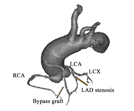
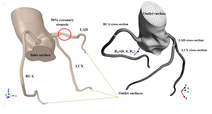
This paper proposes a novel mathematical model of non-Newtonian blood flow and heat transfer in the human coronary system with an external magnetic field. As the blood viscosity is assumed to depend not only on shear rate but also on temperature and magnet strength, the modified Carreau-Yasuda viscosity model is formulated. The computational domain includes the base of the aorta, the right coronary artery, and the left coronary artery, with the left circumflex and left anterior descending arteries. The element-based finite volume method is derived for the solution of the proposed model. Numerical simulations are carried out to investigate the magnetic field effect on the blood flow-heat transfer characteristic in the human coronary system. It is found that the magnetic field has a significant impact on fluid viscosity, leading to enhanced fluid velocity.
Citation: Nattawan Chuchalerm, Wannika Sawangtong, Benchawan Wiwatanapataphee, Thanongchai Siriapisith. Study of Non-Newtonian blood flow - heat transfer characteristics in the human coronary system with an external magnetic field[J]. Mathematical Biosciences and Engineering, 2022, 19(9): 9550-9570. doi: 10.3934/mbe.2022444
This paper proposes a novel mathematical model of non-Newtonian blood flow and heat transfer in the human coronary system with an external magnetic field. As the blood viscosity is assumed to depend not only on shear rate but also on temperature and magnet strength, the modified Carreau-Yasuda viscosity model is formulated. The computational domain includes the base of the aorta, the right coronary artery, and the left coronary artery, with the left circumflex and left anterior descending arteries. The element-based finite volume method is derived for the solution of the proposed model. Numerical simulations are carried out to investigate the magnetic field effect on the blood flow-heat transfer characteristic in the human coronary system. It is found that the magnetic field has a significant impact on fluid viscosity, leading to enhanced fluid velocity.

| [1] |
O. K. Baskurt, H. J. Meiselman, Blood rheology and hemodynamics, Semin. Thromb. Hemostasis, 29 (2003), 435–450. https://doi.org/10.1055/s-2003-44551 doi: 10.1055/s-2003-44551

|
| [2] | G. R. Cokelet, H. J. Meiselman, Macro-and micro-rheological properties of blood, Biomedical and health research-commission of the European communities them IOS press, 69 (2007). |
| [3] | H. A. Baieth, Physical parameters of blood as a non-Newtonian fluid, Int. J. Biomed. Sci., 4 (2008), 323. |
| [4] |
P. Connes, T. Alexy, J. Detterich, M. Romana, M. D. Hardy-Dessources, S. K. Ballas, The role of blood rheology in sickle cell disease, Blood Rev., 30 (2016), 111–118. https://doi.org/10.1016/j.blre.2015.08.005 doi: 10.1016/j.blre.2015.08.005
|
| [5] |
T. Yamamoto, Y. Nagayama, M. Tamura, A blood-oxygenation-dependent increase in blood viscosity due to a static magnetic field, Phys. Med. Biol., 49 (2004), 3267–3277. https://doi.org/10.1088/0031-9155/49/14/017 doi: 10.1088/0031-9155/49/14/017
|
| [6] |
A. Marcinkowska-Gapinska, H. Nawrocka-Bogusz, Analysis of the magnetic field influence on the rheological properties of healthy persons blood, Biomed Res Int., 2013 (2013), 490410. https://doi.org/10.1155/2013/490410 doi: 10.1155/2013/490410
|
| [7] | M. A. Mohaseb, F. A. Shahin, F. M. Ali, H. A. Baieth, Effect of electromagnetic fields on some biomechanical and biochemical properties of rat's blood, Int. J. Mod. Phys.: Conf. Ser., 869 (2017), 012059. |
| [8] |
Y. Haik, V. Pai, C. J. Chen, Apparent viscosity of human blood in a high static magnetic field, J. Magn. Magn. Mater., 225 (2001), 180-186. https://doi.org/10.1016/S0304-8853(00)01249-X doi: 10.1016/S0304-8853(00)01249-X
|
| [9] |
E. E. Tzirtzilakis, A mathematical model for blood flow in magnetic field, Phys. Fluids, 17 (2005), 077103. https://doi.org/10.1063/1.1978807 doi: 10.1063/1.1978807
|
| [10] |
M. A. Ikbal, S. Chakravarty, K. K. Wong, J. Mazumdar, P. K. Mandal, Unsteady response of non-Newtonian blood flow through a stenosed artery in magnetic field, J. Comput. Appl. Math., 230 (2009), 243–259. https://doi.org/10.1016/j.cam.2008.11.010 doi: 10.1016/j.cam.2008.11.010
|
| [11] |
G. Varshney, V. Katiyar, S. Kumar, Effect of magnetic field on the blood flow in artery having multiple stenosis: a numerical study, Int. J. Eng. Sci. Technol., 2 (2010), 967–982. https://doi.org/10.4314/ijest.v2i2.59142 doi: 10.4314/ijest.v2i2.59142
|
| [12] |
R. Ponalagusamy, R. Tamil Selvi, Blood flow in stenosed arteries with radially variable viscosity, peripheral plasma layer thickness and magnetic field, Meccanica, 48 (2013), 2427–2438. https://doi.org/10.1007/s11012-013-9758-z doi: 10.1007/s11012-013-9758-z
|
| [13] |
S. Majee, G. C. Shit, Numerical investigation of MHD flow of blood and heat transfer in a stenosed arterial segment, J. Magn. Magn. Mater., 424 (2017), 137–147. https://doi.org/10.1016/j.jmmm.2016.10.028 doi: 10.1016/j.jmmm.2016.10.028
|
| [14] |
F. Ali, N. A. Sheikh, I. Khan, M. Saqib, Magnetic field effect on blood flow of Casson fluid in axisymmetric cylindrical tube: A fractional model, J. Magn. Magn. Mater., 423 (2017), 327–336. https://doi.org/10.1016/j.jmmm.2016.09.125 doi: 10.1016/j.jmmm.2016.09.125
|
| [15] |
K. Tzirakis, L. Botti, V. Vavourakis, Y. Papaharilaou, Numerical modeling of non-Newtonian biomagnetic fluid flow, Comput. Fluids, 126 (2016), 170–180. https://doi.org/10.1016/j.compfluid.2015.11.016 doi: 10.1016/j.compfluid.2015.11.016
|
| [16] |
K. Gayathri, K. Shailendhra, Mri and blood flow in human arteries: are there any adverse effects?, Cardiovasc. Eng. Technol., 10 (2019), 242–256. https://doi.org/10.1007/s13239-019-00400-x doi: 10.1007/s13239-019-00400-x
|
| [17] |
J. C. Misra, G. C. Shit, H. J. Rath, Flow and heat transfer of a MHD viscoelastic fluid in a channel with stretching walls: Some applications to haemodynamics, Comput. Fluids, 37 (2008), 1–11. https://doi.org/10.1016/j.compfluid.2006.09.005 doi: 10.1016/j.compfluid.2006.09.005
|
| [18] |
A. A. Dar, Effect of thermal radiation, temperature jump and inclined magnetic field on the peristaltic transport of blood flow in an asymmetric channel with variable viscosity and heat absorption/generation, Iran. J. Sci. Technol., 45 (2021), 487–501. https://doi.org/10.1007/s40997-020-00349-6 doi: 10.1007/s40997-020-00349-6
|
| [19] |
H. Alimohamadi, K. Sadeghy, On the use of magnetic fields for controlling the temperature of hot spots on porous plaques in stenosis arteries, J. Soc. Rheol., Jpn., 43 (2016), 135–144. https://doi.org/10.1678/rheology.43.135 doi: 10.1678/rheology.43.135
|
| [20] |
S. Nadeem, N. S. Akbar, Peristaltic flow of a Jeffrey fluid with variable viscosity in an asymmetric channel, Z. Naturforsch. A, 64 (2009), 713–722. https://doi.org/10.1515/zna-2009-1107 doi: 10.1515/zna-2009-1107
|
| [21] |
O. D. Makinde, O. O. Onyejekwe, A numerical study of MHD generalized Couette flow and heat transfer with variable viscosity and electrical conductivity, J. Magn. Magn. Mater., 323 (2011), 2757–2763. https://doi.org/10.1016/j.jmmm.2011.05.040 doi: 10.1016/j.jmmm.2011.05.040
|
| [22] |
G. C. Shit, S. Majee, Pulsatile flow of blood and heat transfer with variable viscosity under magnetic and vibration environment, J. Magn. Magn. Mater., 388 (2015), 106–115. https://doi.org/10.1016/j.jmmm.2015.04.026 doi: 10.1016/j.jmmm.2015.04.026
|
| [23] |
R. Tao, K. Huang, K. Reducing blood viscosity with magnetic fields, Phys. Rev. E, 84 (2011), 011905. https://doi.org/10.1103/PhysRevE.84.011905 doi: 10.1103/PhysRevE.84.011905
|
| [24] | A. A. Kadhim, B. T. Seah, A. M. Zubair, Influence of magnetic field on blood viscosity, Adv. Environ. Biol., 10 (2016), 107–111. |
| [25] | A. Keshtkar, M. Roozbeh, H. K. Sani, Fluctuation in blood velocity under applied electromagnetic pulse fields, Int. J. Biosen. Bioelectron., 2 (2017), 107–112. |
| [26] |
A. Javadzadegan, A. Moshfegh, M. Behnia, Effect of magnetic field on haemodynamic perturbations in atherosclerotic coronary arteries, J. Med. Eng. Technol., 42 (2018), 148–156. https://doi.org/10.1080/03091902.2018.1447034 doi: 10.1080/03091902.2018.1447034
|
| [27] |
Y. Zare, S. P. Park, K. Y. Rhee, Analysis of complex viscosity and shear thinning behavior in poly (lactic acid)/poly (ethylene oxide)/carbon nanotubes biosensor based on Carreau–Yasuda model, Results Phys., 13 (2019), 102245. https://doi.org/10.1016/j.rinp.2019.102245 doi: 10.1016/j.rinp.2019.102245
|
| [28] |
A. Sharma, P. Kumar, U. Gupta, The effect of magnetic-field-dependent viscosity and rotation on ferrothermohaline convection saturating a porous medium in the presence of dust particles, J. Geophys. Eng., 2 (2005), 238. https://doi.org/10.1088/1742-2132/2/3/008 doi: 10.1088/1742-2132/2/3/008
|
| [29] | M. G. ibrahim, Numerical simulation to the activation energy study on blood flow of seminal nanofluid with mixed convection effects, Comput. Methods Biomech. Biomed. Eng., (2022), 1–11. https://doi.org/10.1080/10255842.2022.206301 |
| [30] |
M. Sankaranarayanan, L. P. Chua, D. N. Ghista, Y. S. Tan, Computational model of blood flow in the aorto-coronary bypass graft, BioMed. Eng., 4 (2005), 1–13. https://doi.org/10.1186/1475-925X-4-14 doi: 10.1186/1475-925X-4-14
|
| [31] |
H. J. Kim, I. E. Vignon-Clementel, J. S. Coogan, C. A. Figueroa, K. E. Jansen, C. A. Taylor, Patient-specific modeling of blood flow and pressure in human coronary arteries, Ann. Biomed. Eng., 38 (2010), 3195–3209. https://doi.org/10.1007/s10439-010-0083-6 doi: 10.1007/s10439-010-0083-6
|
| [32] | E. W. Lo, L. J. Menezes, R. Torii, Impact of inflow boundary conditions on the calculation of CT-based FFR Fluids, 4 (2019), 60. https://doi.org/10.3390/fluids4020060 |
| [33] |
B. Wiwatanapataphee, Y. H. Wu, T. Siriapisith, B. Nuntadilok, Effect of branchings on blood flow in the system of human coronary arteries, Math. Biosci. Eng., 9 (2012), 199–214. https://doi.org/10.3934/mbe.2012.9.199 doi: 10.3934/mbe.2012.9.199
|
| [34] | P. A. Hasgall, F. Di Gennaro, C. Baumgartner, E. Neufeld, B. Lloyd, M. C. Gosselin, et al,, IT'IS Database for Thermal and Electromagnetic Parameters of Biological Tissues, Version 4.1, IT'IS Found., (2022). https://doi.org/10.13099/VIP21000-04-1 |
| [35] |
Z. Tyfa, D. Obidowski, P. Reorowicz, L. Stefańczyk, J. Fortuniak, K. Jóźwik, Numerical simulations of the pulsatile blood flow in the different types of arterial fenestrations: Comparable analysis of multiple vascular geometries, Biocybernetics and Biomedical Engineering, 38 (2018), 228–242. https://doi.org/10.1016/j.bbe.2018.01.004 doi: 10.1016/j.bbe.2018.01.004
|
| [36] |
J. Biasetti, F. Hussain, T. C. Gasser, Blood flow and coherent vortices in the normal and aneurysmatic aortas: a fluid dynamical approach to intra-luminal thrombus formation, J. R. Soc. Interface, 63 (2011), 1449–1461. https://doi.org/10.1098/rsif.2011.0041 doi: 10.1098/rsif.2011.0041
|
| [37] | ADAM editorial team, Body temperature norms: Medlineplus medical encyclopedia, 2018. Available from: https://medlineplus.gov/ency/article/001982.htm. |
| [38] |
K. S. Sakariassen, L. Orning, V. T. Turitto, The impact of blood shear rate on arterial thrombus formation, Future Sci. OA, 1 (2015), FSO30. https://doi.org/10.4155/fso.15.28 doi: 10.4155/fso.15.28
|
Figures(11) / Tables(4)

Nattawan Chuchalerm, Wannika Sawangtong, Benchawan Wiwatanapataphee, Thanongchai Siriapisith. Study of Non-Newtonian blood flow - heat transfer characteristics in the human coronary system with an external magnetic field[J]. Mathematical Biosciences and Engineering, 2022, 19(9): 9550-9570. doi: 10.3934/mbe.2022444


 DownLoad: