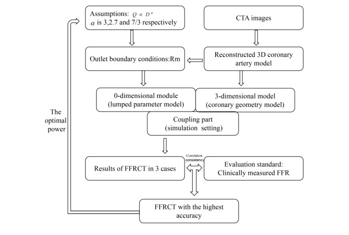
To explore the influence of the blood flow-diameter scaling laws of $ \mathrm{Q}\mathrm{\alpha }{\mathrm{D}}^{3} $, $ \mathrm{Q}\mathrm{\alpha }{\mathrm{D}}^{2.7} $ and $ \text{Q}\alpha \text{D}{}^{7}\!\!\diagup\!\!{}_{3}\; $ on the numerical simulation of fraction flow reserve based on CTA images and to find the optimal exponents.
1) 26 patients with coronary artery disease were screened according to the inclusion criteria; 2) Microcirculation resistance (Rm) was calculated under the 3, 2.7 and 7/3 power of the flow-diameter scaling law, which were recorded as 3Rm, 2.7Rm and 7/3Rm, respectively; 3) 3Rm, 2.7Rm and 7/3Rm were used as exit boundary conditions to simulate FFRCT, quoted as 3FFRCT, 2.7FFRCT and 7/3FFRCT, respectively; 4) The correlation and diagnostic performance between three kinds of FFRCT and FFR were analyzed.
The p-values of comparing 3Rm, 2.7Rm and 7/3Rm with FFR were 0.004, 0.005 and 0.010, respectively; the r value between 7/3FFRCT and FFR (0.96) was better than that of 3FFRCT (0.95) and 2.7FFRCT (0.95); the 95% LoA between 7/3FFRCT and FFR (-0.08~0.11) was smaller than that of 3FFRCT (-0.10~0.12) and 2.7FFRCT (-0.09~0.11); the AUC and accuracy of 7/3FFRCT [0.962 (0.805-0.999), 96.15%] were the same as those of 2.7FFRCT [0.962 (0.805-0.999), 96.15%] and better than those of 3FFRCT [0.944 (0.777-0.996), 92.3%]. The prediction threshold of 7/3FFRCT (0.791) was closer to 0.8 than that of 3FFRCT (0.816) and 2.7FFRCT (0.787).
The blood flow-diameter scaling law affects the FFRCT simulation by influencing the exit boundary condition Rm of the calculation. With $ Q\alpha D{}^{7}\!\!\diagup\!\!{}_{3}\; $, FFRCT had the highest diagnostic performance. The blood flow-diameter scaling law provides theoretical support for the blood flow distribution in the bifurcated vessel and improves the FFRCT model.
Citation: Na Li, Bao Li, Yili Feng, Junling Ma, Liyuan Zhang, Jian Liu, Youjun Liu. Impact of coronary bifurcated vessels flow-diameter scaling laws on fractional flow reserve based on computed tomography images (FFRCT)[J]. Mathematical Biosciences and Engineering, 2022, 19(3): 3127-3146. doi: 10.3934/mbe.2022145
To explore the influence of the blood flow-diameter scaling laws of $ \mathrm{Q}\mathrm{\alpha }{\mathrm{D}}^{3} $, $ \mathrm{Q}\mathrm{\alpha }{\mathrm{D}}^{2.7} $ and $ \text{Q}\alpha \text{D}{}^{7}\!\!\diagup\!\!{}_{3}\; $ on the numerical simulation of fraction flow reserve based on CTA images and to find the optimal exponents.
1) 26 patients with coronary artery disease were screened according to the inclusion criteria; 2) Microcirculation resistance (Rm) was calculated under the 3, 2.7 and 7/3 power of the flow-diameter scaling law, which were recorded as 3Rm, 2.7Rm and 7/3Rm, respectively; 3) 3Rm, 2.7Rm and 7/3Rm were used as exit boundary conditions to simulate FFRCT, quoted as 3FFRCT, 2.7FFRCT and 7/3FFRCT, respectively; 4) The correlation and diagnostic performance between three kinds of FFRCT and FFR were analyzed.
The p-values of comparing 3Rm, 2.7Rm and 7/3Rm with FFR were 0.004, 0.005 and 0.010, respectively; the r value between 7/3FFRCT and FFR (0.96) was better than that of 3FFRCT (0.95) and 2.7FFRCT (0.95); the 95% LoA between 7/3FFRCT and FFR (-0.08~0.11) was smaller than that of 3FFRCT (-0.10~0.12) and 2.7FFRCT (-0.09~0.11); the AUC and accuracy of 7/3FFRCT [0.962 (0.805-0.999), 96.15%] were the same as those of 2.7FFRCT [0.962 (0.805-0.999), 96.15%] and better than those of 3FFRCT [0.944 (0.777-0.996), 92.3%]. The prediction threshold of 7/3FFRCT (0.791) was closer to 0.8 than that of 3FFRCT (0.816) and 2.7FFRCT (0.787).
The blood flow-diameter scaling law affects the FFRCT simulation by influencing the exit boundary condition Rm of the calculation. With $ Q\alpha D{}^{7}\!\!\diagup\!\!{}_{3}\; $, FFRCT had the highest diagnostic performance. The blood flow-diameter scaling law provides theoretical support for the blood flow distribution in the bifurcated vessel and improves the FFRCT model.

| [1] |
N. H. J. Pijls, B. DeBruyne, K. Peels, P. H. VanderVoort, H. Bonnier, J. Bartunek, et al., Measurement of fractional flow reserve to assess the functional severity of coronary-artery stenoses, N. Engl. J. Med., 334 (1996), 1703-1708. https://doi.org/10.1056/NEJM199606273342604 doi: 10.1056/NEJM199606273342604

|
| [2] |
Y. Feng, B. Y. Mao, B. Li, J. Liu, J. C. Liu, Y. J. Liu, Effect of hemodynamic parameters on fractional flow reserve, J. Mech. Med. Biol., 20 (2020), 14. https://doi.org/10.1142/S0219519420500177 doi: 10.1142/S0219519420500177
|
| [3] |
N. Kakouros, F. J. Rybicki, D. Mitsouras, J. M. Miller, Coronary pressure-derived fractional flow reserve in the assessment of coronary artery stenoses, Eur. Radiol., 23 (2013), 958-967. https://doi.org/10.1007/s00330-012-2670-4 doi: 10.1007/s00330-012-2670-4
|
| [4] |
C. Ball, G. Pontone, M. Rabbat, Fractional flow reserve derived from coronary computed tomography angiography datasets: The next frontier in noninvasive assessment of coronary artery disease, Biomed. Res. Int., (2018), 8. https://doi.org/10.1155/2018/2680430 doi: 10.1155/2018/2680430
|
| [5] |
S. X. Tu, J. Westra, J. Adjedj, D. X. Ding, F. Y. Liang, B. Xu, et al., Fractional flow reserve in clinical practice: from wire-based invasive measurement to image-based computation, Eur. Heart J., 41 (2020), 3271-3279. https://doi.org/10.1093/eurheartj/ehz918 doi: 10.1093/eurheartj/ehz918
|
| [6] |
M. T. Lu, M. Ferencik, R. S. Roberts, K. L. Lee, A. Ivanov, E. Adami, et al., Noninvasive FFR derived from coronary CT angiography management and outcomes in the PROMISE trial, JACC Cardiovasc. Imaging, 10 (2017), 1350-1358. https://doi.org/10.1016/j.jcmg.2016.11.024 doi: 10.1016/j.jcmg.2016.11.024
|
| [7] |
B. K. Koo, A. Erglis, J. H. Doh, D. V. Daniels, S. Jegere, H. S. Kim, et al., Diagnosis of ischemia-causing coronary stenoses by noninvasive fractional flow reserve computed from coronary computed tomographic angiograms. Results from the prospective multicenter DISCOVER-FLOW (Diagnosis of Ischemia-Causing Stenoses Obtained Via Noninvasive Fractional Flow Reserve) Study, J. Am. Coll. Cardiol., 58 (2011), 1989-1997. https://doi.org/10.1016/j.jacc.2011.06.066 doi: 10.1016/j.jacc.2011.06.066
|
| [8] | J. K. Min, D. S. Berman, M. J. Budoff, F. A. Jaffer, J. Leipsic, M. B. Leon, et al., Rationale and design of the DeFACTO (Determination of Fractional Flow Reserve by Anatomic Computed Tomographic AngiOgraphy) study, J. Cardiovasc. Comput. Tomogr., 5 (2011), 301-309. |
| [9] |
C. A. Taylor, T. A. Fonte, J. K. Min, Computational fluid dynamics applied to cardiac computed tomography for noninvasive quantification of fractional flow reserve scientific basis, JACC, 61 (2013), 2233-2241. https://doi.org/10.1016/j.jacc.2012.11.083 doi: 10.1016/j.jacc.2012.11.083
|
| [10] |
H. J. Kim, I. E. Vignon-Clementel, J. S. Coogan, C. A. Figueroa, K. E. Jansen, C. A. Taylor, Patient-specific modeling of blood flow and pressure in human coronary arteries, Ann. Biomed. Eng., 38 (2010), 3195-3209. https://doi.org/10.1007/s10439-010-0083-6 doi: 10.1007/s10439-010-0083-6
|
| [11] |
C. K. Zarins, C. A. Taylor, J. K. Min, Computed fractional flow reserve (FFTCT) derived from coronary CT angiography, J. Cardiovasc. Transl. Res., 6 (2013), 708-714. https://doi.org/10.1007/s12265-013-9498-4 doi: 10.1007/s12265-013-9498-4
|
| [12] |
M. Gotberg, E. H. Christiansen, I. J. Gudmundsdottir, L. Sandhall, M. Danielewicz, L. Jakobsen, et al., Instantaneous wave-free ratio versus fractional flow reserve to guide PCI, N. Engl. J. Med., 376 (2017), 1813-1823. https://doi.org/10.1056/NEJMoa1616540 doi: 10.1056/NEJMoa1616540
|
| [13] |
Y. P. van de Hoef, F. Nolte, P. Damman, R. Delewi, M. Bax, S. A. J. Chamuleau, et al., Diagnostic accuracy of combined intracoronary pressure and flow velocity information during baseline conditions adenosine-free assessment of functional coronary lesion severity, Circ. Cardiovasc. Interv., 5 (2012), 508-514. https://doi.org/10.1161/CIRCINTERVENTIONS.111.965707 doi: 10.1161/CIRCINTERVENTIONS.111.965707
|
| [14] |
G. S. Kassab, J. Berkley, Y. C. B. Fung, Analysis of pig's coronary arterial blood flow with detailed anatomical data, Ann. Biomed. Eng., 25 (1997), 204-217. https://doi.org/10.1007/BF02738551 doi: 10.1007/BF02738551
|
| [15] |
G. S. Kassab, Y. C. B. Fung, The pattern of coronary arteriolar bifurcations and the uniform shear hypothesis, Ann. Biomed. Eng., 23 (1995), 13-20. https://doi.org/10.1007/bf02368296 doi: 10.1007/bf02368296
|
| [16] |
G. S. Kassab, C. A. Rider, N. J. Tang, Y. C. B. Fung, Morphometry of pig coronary arterial trees, Am. J. Physiol., 265 (1993), 350-365. https://doi.org/10.1152/ajpheart.1993.265.1.H350 doi: 10.1152/ajpheart.1993.265.1.H350
|
| [17] |
Y. L. Huo, G. S. Kassab, Intraspecific scaling laws of vascular trees, J. R. Soc. Interface, 9 (2012), 190-200. https://doi.org/10.1098/rsif.2011.0270 doi: 10.1098/rsif.2011.0270
|
| [18] |
T. F. Sherman, On connecting large vessels to small-the meaning of murray law, J. Gen. Physiol., 78 (1981), 431-453. https://doi.org/10.1085/jgp.78.4.431 doi: 10.1085/jgp.78.4.431
|
| [19] |
Y. F. Zhou, G. S. Kassab, S. Molloi, On the design of the coronary arterial tree: a generalization of Murray's law, Phys. Med. Biol., 44 (1999), 2929-2945. https://doi.org/10.1088/0031-9155/44/12/306 doi: 10.1088/0031-9155/44/12/306
|
| [20] |
G. S. Kassab, Scaling laws of vascular trees: of form and function, Am. J. Physiol. Heart Circ. Physiol., 290 (2006), H894-H903. https://doi.org/10.1152/ajpheart.00579.2005 doi: 10.1152/ajpheart.00579.2005
|
| [21] |
L. Itu, P. Sharma, C. Suciu, F. Moldoveanu, D. Comaniciu, Personalized blood flow computations: A hierarchical parameter estimation framework for tuning boundary conditions, Int. J. Numer. Method Biomed. Eng., 33 (2017), e02823. https://doi.org/10.1002/cnm.2803 doi: 10.1002/cnm.2803
|
| [22] |
J. P. H. M. van den Wijngaard, J. C. V. Schwarz, P. van Horssen, M. van Lier, J. G. G. Dobbe, J. A. E. Spaan, et al., 3D Imaging of vascular networks for biophysical modeling of perfusion distribution within the heart, J. Biomech., 46 (2013), 229-239. https://doi.org/10.1016/j.jbiomech.2012.11.027 doi: 10.1016/j.jbiomech.2012.11.027
|
| [23] |
J. M. Zhang, T. Luo, S. Y. Tan, A. M. Lomarda, A. S. L. Wong, F. Y. J. Keng, et al., Hemodynamic analysis of patient-specific coronary artery tree, Int. J. Numer. Method Biomed. Eng., 31 (2015), e02708. https://doi.org/10.1002/cnm.2708 doi: 10.1002/cnm.2708
|
| [24] |
M. Rabbat, J. Leipsic, J. Bax, B. Kauh, R. Verma, D. Doukas, et al., Fractional flow reserve derived from coronary computed tomography angiography safely defers invasive coronary angiography in patients with stable coronary artery disease, J. Clin. Med., 9 (2020), 15. https://doi.org/10.3390/jcm9020604 doi: 10.3390/jcm9020604
|
| [25] |
J. K. Min, J. Leipsic, M. J. Pencina, D. S. Berman, B. K. Koo, C. van Mieghem, et al., Diagnostic accuracy of fractional flow reserve from anatomic CT angiography, JAMA, 308 (2012), 1237-1245. https://doi.org/10.1001/2012.jama.11274 doi: 10.1001/2012.jama.11274
|
| [26] | A. Wahle, E. Wellnhofer, I. Mugaragu, H. U. Sauer, H. Oswald, E. Fleck, Quantitative volume analysis of coronary vessel systems by 3-D reconstruction from biplane angiograms, in 1993 IEEE Conference Record Nuclear Science Symposium and Medical Imaging Conference, 3 (1993), 1217-1221. https://doi.org/10.1109/NSSMIC.1993.701838 |
| [27] |
B. Y. Mao, W. X. Wang, Z. Zhao, X. Zhao, L. L. Li, H. X. Zhang, et al., On the relationship between competitive flow and FFT analysis of the flow waves in the left internal mammary artery graft in the process of CABG, Biomed. Eng. Online, 15 (2016), 557-567. https://doi.org/10.1186/s12938-016-0260-4 doi: 10.1186/s12938-016-0260-4
|
| [28] |
G. B. West, J. H. Brown, B. J. Enquist, The fourth dimension of life: Fractal geometry and allometric scaling of organisms, Science, 284 (1999), 1677-1679. https://doi.org/10.1126/science.284.5420.1677 doi: 10.1126/science.284.5420.1677
|
| [29] |
C. Tesche, K. Otani, C. N. de Cecco, A. Coenen, J. De Geer, M. Kruk, et al., Influence of coronary calcium on diagnostic performance of machine learning CT-FFR results from machine registry, JACC Cardiovasc. Imaging, 13 (2020), 760-770. https://doi.org/10.1016/j.jcmg.2019.06.027 doi: 10.1016/j.jcmg.2019.06.027
|
| [30] | J. K. Min, D. Berman, L. J. Shaw, L. Mauri, B. K. Koo, C. van Mieghem, et al., Fractional flow reserved derived from computed tomographic angiography (FFRCT) to discriminate individuals with versus without Ischemia: Results from the DeFACTO trial (determination of fractional flow reserve by anatomic computed tomographic angiography), Circulation, 126 (2012). |
| [31] |
J. Escaned, M. Echavarria-Pinto, H. M. Garcia-Garcia, T. P. van de Hoef, T. de Vries, P. Kaul, et al., Prospective assessment of the diagnostic accuracy of instantaneous wave-free ratio to assess coronary stenosis relevance: results of ADVISE Ⅱ International, Multicenter Study (ADenosine Vasodilator Independent Stenosis Evaluation Ⅱ), JACC Cardiovasc. Interv., 8 (2015), 824-833. https://doi.org/10.1016/j.jcin.2015.01.029 doi: 10.1016/j.jcin.2015.01.029
|
| [32] |
L. Itu, S. Rapaka, T. Passerini, B. Georgescu, C. Schwemmer, M. Schoebinger, et al., A machine-learning approach for computation of fractional flow reserve from coronary computed tomography, J. Appl. Physiol., 121 (2015), 42-52. https://doi.org/10.1152/japplphysiol.00752.2015 doi: 10.1152/japplphysiol.00752.2015
|
| [33] |
K. Greff, R. K. Srivastava, J. Koutník, B. R. Steunebrink, J. Schmidhuber, LSTM: a search space odyssey, IEEE Trans. Neural Networks Learn. Syst., 28 (2017), 2222-2232. https://doi.org/10.1109/TNNLS.2016.2582924 doi: 10.1109/TNNLS.2016.2582924
|
| [34] |
Z. Gao, Y. Li, Y. Sun, J. Yang, H. Xiong, H. Zhang, et al., Motion tracking of the carotid artery wall from ultrasound image sequences: a nonlinear state-space approach, IEEE Trans. Med. Imaging, 37 (2018), 273-283. https://doi.org/10.1109/TMI.2017.2746879 doi: 10.1109/TMI.2017.2746879
|
| [35] |
Z. Gao, X. Liu, S. Qi, W. Wu, W. K. Hau, H. Zhan, Automatic segmentation of coronary tree in CT angiography images, Int. J. Adapt. Control Signal Process., 33 (2019), 1239-1247. https://doi.org/10.1002/acs.2762 doi: 10.1002/acs.2762
|
| [36] |
Z. Gao, X. Wang, S. Sun, D. Wu, J. Bai, Y. Yin, et al., Learning physical properties in complex visual scenes: An intelligent machine for perceiving blood flow dynamics from static CT angiography imaging, Neural Networks, 123 (2020), 82-93. https://doi.org/10.1016/j.neunet.2019.11.017 doi: 10.1016/j.neunet.2019.11.017
|
| [37] |
P. K. Siogkas, L. Lakkas, A. I. Sakellarios, G. Rigas, S. Kyriakidis, K. A. Stefanou, et al., SmartFFR, a new functional index of coronary stenosis: comparison with invasive ffr data, Front. Cardiovasc. Med., (2021), 958. https://doi.org/10.3389/fcvm.2021.714471 doi: 10.3389/fcvm.2021.714471
|


Figures(10) / Tables(2)

Na Li, Bao Li, Yili Feng, Junling Ma, Liyuan Zhang, Jian Liu, Youjun Liu. Impact of coronary bifurcated vessels flow-diameter scaling laws on fractional flow reserve based on computed tomography images (FFRCT)[J]. Mathematical Biosciences and Engineering, 2022, 19(3): 3127-3146. doi: 10.3934/mbe.2022145


 DownLoad: