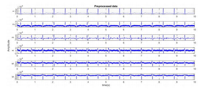
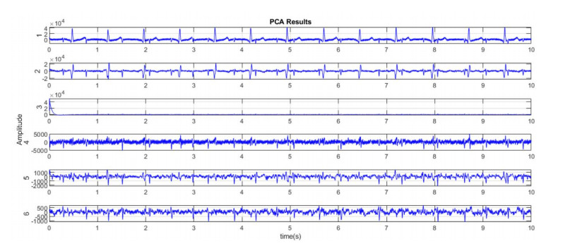
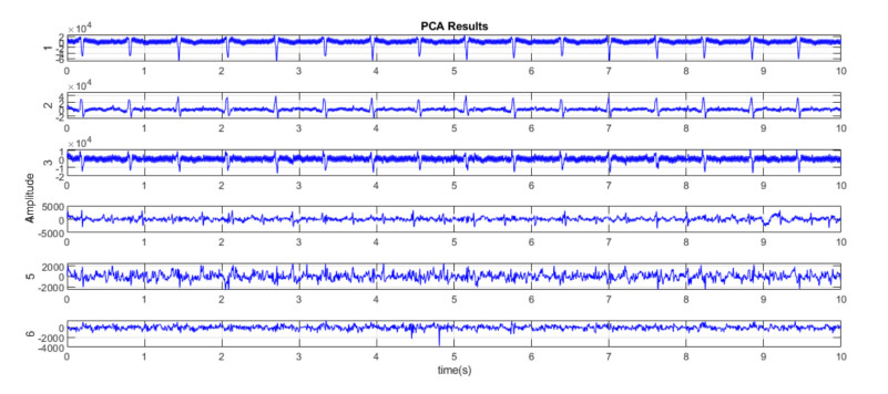
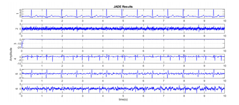
The fetal heart rate (fHR) variability and fetal electrocardiogram (fECG) are considered the most important sources of information about fetal wellbeing. Non-invasive fetal monitoring and analysis of fECG are paramount for clinical trials. They enable examining the fetal health status and detecting the heart rate changes associated with insufficient oxygenation to cut the likelihood of hypoxic fetal injury. Despite the fact that significant advances have been achieved in electrocardiography and adult ECG signal processing, the analysis of fECG is still in its infancy. Due to accurate fetal morphology extraction techniques have not been properly developed, many areas require particular attention on the way of fully understanding the changes in variability in the fetus and implementation of the non-invasive techniques suitable for remote home care which is increasingly in demand for high-risk pregnancy monitoring. In this paper, we introduce an integrated approach for fECG signal extraction and processing based on various methods for fetal welfare investigation and hypoxia risk estimation. To the best of our knowledge, this is the first attempt to introduce the auto-generated risk scoring in fECG to achieve early warning on fetus' safety and provide the physician with additional information about the possible fetal complications. The proposed method includes the following stages: fECG extraction, fHR and fetal heart rate variability (fHRV) calculation, hypoxia index (HI) evaluation and risk estimation. The extracted signals were examined by assessing Signal to Noise Ratio (SNR) and mean square error (MSE) values. The results obtained demonstrated great potential, but more profound research and validation, as well as a consistent clinical study, are needed before implementation into the hospital and at-home monitoring.
Citation: Tetiana Biloborodova, Lukasz Scislo, Inna Skarga-Bandurova, Anatoliy Sachenko, Agnieszka Molga, Oksana Povoroznyuk, Yelyzaveta Yevsieieva. Fetal ECG signal processing and identification of hypoxic pregnancy conditions in-utero[J]. Mathematical Biosciences and Engineering, 2021, 18(4): 4919-4942. doi: 10.3934/mbe.2021250
The fetal heart rate (fHR) variability and fetal electrocardiogram (fECG) are considered the most important sources of information about fetal wellbeing. Non-invasive fetal monitoring and analysis of fECG are paramount for clinical trials. They enable examining the fetal health status and detecting the heart rate changes associated with insufficient oxygenation to cut the likelihood of hypoxic fetal injury. Despite the fact that significant advances have been achieved in electrocardiography and adult ECG signal processing, the analysis of fECG is still in its infancy. Due to accurate fetal morphology extraction techniques have not been properly developed, many areas require particular attention on the way of fully understanding the changes in variability in the fetus and implementation of the non-invasive techniques suitable for remote home care which is increasingly in demand for high-risk pregnancy monitoring. In this paper, we introduce an integrated approach for fECG signal extraction and processing based on various methods for fetal welfare investigation and hypoxia risk estimation. To the best of our knowledge, this is the first attempt to introduce the auto-generated risk scoring in fECG to achieve early warning on fetus' safety and provide the physician with additional information about the possible fetal complications. The proposed method includes the following stages: fECG extraction, fHR and fetal heart rate variability (fHRV) calculation, hypoxia index (HI) evaluation and risk estimation. The extracted signals were examined by assessing Signal to Noise Ratio (SNR) and mean square error (MSE) values. The results obtained demonstrated great potential, but more profound research and validation, as well as a consistent clinical study, are needed before implementation into the hospital and at-home monitoring.

| [1] |
C. Gravett, L. O. Eckert, M. G. Gravett, D. J. Dudley, E. M. Stringer, T. B. M. Mujobu, et al., Non-reassuring fetal status: case definition & guidelines for data collection, analysis, and presentation of immunization safety data, Vaccine, 34 (2016), 6084. doi: 10.1016/j.vaccine.2016.03.043

|
| [2] |
J. Fahey, T. L. King, Intrauterine asphyxia: clinical implications for providers of intrapartum care, J. Midw. Womens Health, 50 (2005), 498-506. doi: 10.1016/j.jmwh.2005.08.007
|
| [3] | R. K. Freeman, T. J. Garite, M. P. Nageotte, L. A. Miller, Fetal Heart Rate Monitoring, 4nd edition, Philadelphia, Lippincott Williams and Wilkins, 2012. |
| [4] | GE Healthcare, Novii Wireless Patch System, Available from: https://www.gehealthcare.com/products/maternal-infant-care/fetal-monitors/novii-wireless-patch-system. |
| [5] |
M. Mhajna, N. Schwartz, L. Levit-Rosen, S. Warsof, M. Lipschuetz, M. Jakobs, et al., Wireless, remote solution for home fetal and maternal heart rate monitoring, Am. J. Obstet. Gynecol. MFM, 2 (2020), 100101. doi: 10.1016/j.ajogmf.2020.100101
|
| [6] | R. Tapia-Conyer, S. Lyford, R. Saucedo, M. Casale, H. Gallardo, K. Becerra, et al., Improving perinatal care in the rural regions worldwide by wireless enabled antepartum fetal monitoring: a demonstration project, Int. J. Telemed. Appl., 2015. |
| [7] |
E. M. Graatsma, B. C. Jacod, L. A. J. V. Egmond, E. J. H. Mulder, G. H. A. Visser, Fetal electrocardiography: feasibility of long-term fetal heart rate recordings, Bjog, 116 (2009), 334-338. doi: 10.1111/j.1471-0528.2008.01951.x
|
| [8] |
R. Martinek, R. Kahankova, B. Martin, J. Nedoma, M. Fajkus, A novel modular fetal ECG STAN and HRV analysis: towards robust hypoxia detection, J. Health Care Technol., 27 (2019), 257-287. doi: 10.3233/THC-181375
|
| [9] |
J. Balayla, G. Shrem, Use of artificial intelligence (AI) in the interpretation of intrapartum fetal heart rate (FHR) tracings: a systematic review and meta-analysis, Arch Gynecol. Obstet., 300 (2019), 7-14. doi: 10.1007/s00404-019-05151-7
|
| [10] |
G. M. Ungureanu, J. W. M. Bergmans, S. G. Oei, A. Ungureanu, W. Wolf, The event synchronous canceller algorithm removes maternal ECG from abdominal signals without affecting the fetal ECG, Comput. Biol. Med., 39 (2009), 562-567. doi: 10.1016/j.compbiomed.2009.03.013
|
| [11] | A. Agostinelli, S. Fioretti, F. D. Nardo, L. Burattini, Clinical application of the segmented-beat modulation method for fetal ECG extraction, in Proceedings of the 12th International Workshop on Intelligent Solutions in Embedded Systems (WISES), Ancona, IEEE Press, (2015), 35-40. |
| [12] |
I. Skarga-Bandurova, T. Biloborodova, M. Nesterov, Extracting interesting rules from gestation course data for early diagnosis of neonatal hypoxia, J. Med. Syst., 43 (2019), 1-10. doi: 10.1007/s10916-018-1115-2
|
| [13] | R. Sameni, G. D. Clifford, A review of fetal ECG signal processing; issues and promising directions, Open Pacing, Electrophysiol. Ther. J., 3 (2010), 4-20. |
| [14] | D. Taralunga, M. G. Ungureanu, I. Gussi, R. Strungaru, W. Wolf, Fetal ECG extraction from abdominal signals: a review on suppression of fundamental power line interference component and its harmonics, Comput. Math. Method. Med., (2014), 239060. |
| [15] |
J. Jezewski, A. Matonia, T. Kupka, D. Roj, R. Czabanski, Determination of the fetal heart rate from abdominal signals: evaluation of beat-to-beat accuracy in relation to the direct fetal electrocardiogram, Biomed. Eng., 57 (2012), 383-394. doi: 10.1515/bmt-2012-4501
|
| [16] |
J. Behar, A. Johnson, G. D. Clifford, J. Oster, A comparison of single channel fetal ECG extraction methods, Ann. Biomed. Eng., 42 (2014), 1340-1353. doi: 10.1007/s10439-014-0993-9
|
| [17] |
D. Jagannath, A. I. Selvakumar, Issues and research on foetal electrocardiogram signal elicitation, Biomed. Signal Process. Control, 10 (2014), 224-244. doi: 10.1016/j.bspc.2013.11.001
|
| [18] | S. Ravindrakumar, K. B. Raja, Fetal ECG extraction and enhancement in prenatal monitoring-Review and implementation issues, in Proceedings of the IEEE Trendz in Information Sciences & Computing (TISC), Chennai, India, (2010), 16-20. |
| [19] |
N. Widatalla, Y. Kasahara, Y. Kimura, A. Khandoker, Model based estimation of QT intervals in non-invasive fetal ECG signals, Plos one, 15 (2020), e0232769. doi: 10.1371/journal.pone.0232769
|
| [20] |
Q. Yu, H. Yan, L. Song, W. Guo, H. Liu, J. Si, et al., Automatic identifying of maternal ECG source when applying ICA in fetal ECG extraction, Biocybern. Biomed. Eng., 38 (2018), 448-455. doi: 10.1016/j.bbe.2018.03.003
|
| [21] | S. Padhy, Mult acne monitoring bioimpedance deviceilead ECG data analysis using SVD and higher-order SVD, Doctoral dissertation, Ph. D. thesis, Indian Institute of Technology Guwahati, India, 2017. |
| [22] | A. D. Deogire, Multi lead fetal QRS detection with principal component analysis, in IEEE 2018 3rd International Conference for Convergence in Technology (I2CT), (2018), 1-5. |
| [23] | I. Perova, Y. Bodyanskiy, Adaptive human machine interaction approach for feature selection-extraction task in medical data mining, IJC 17 (2018), 113-119. |
| [24] | M. Fatemi, R. Sameni, An online subspace denoising algorithm for maternal ECG removal from fetal ECG signals, IJST-T Electr. Eng., 41 (2017), 65-79. |
| [25] | Z. L. Zhang, Y. Ye, Extended Barros's extraction algorithm with its application in fetal ECG extraction, in 2005 International Conference on Neural Networks and Brain, Beijing, (2005), 1077-1080. |
| [26] |
S. Redif, Fetal electrocardiogram estimation using polynomial eigenvalue decomposition, Turk. J. Electr. Eng. Comput. Sci., 24 (2016), 2483-2497. doi: 10.3906/elk-1401-19
|
| [27] | G. H. Golub, C. F. van Loan, Matrix Computations, 3nd edition, Baltimore, The Johns Hopkins University Press, 1996. |
| [28] | M. Suganthy, Analysis of R-peaks in fetal electrocardiogram to detect heart disorder using fuzzy clustering, in IEEE 5th International Conference for Convergence in Technology (I2CT), (2019), 1-4. |
| [29] |
N. E. Huang, Z. Wu, S. R. Long, Hilbert-Huang transform, Scholarpedia J., 3 (2008), 2544. doi: 10.4249/scholarpedia.2544
|
| [30] |
R. Jaros, R. Martinek, R. Kahankova, Non-adaptive methods for fetal ECG signal processing: a review and appraisal, Sensors, 18 (2018), 3648. doi: 10.3390/s18113648
|
| [31] |
A. Hyvarinen, Fast and robust fixed-point algorithms for independent component analysis, IEEE Trans. Neural Netw., 10 (1999), 626-634. doi: 10.1109/72.761722
|
| [32] | J. F. Cardoso, Source separation using higher order moments, in Proceedings of the IEEE International Conference on Accoustics, Speech and Signal Processing, (1989), 2109-2112. |
| [33] |
L. Yuan, Y. Yuan, Z. Zhou, Y. Bai, S. Wu, A fetal ECG monitoring system based on the Android smartphone, Sensors, 19 (2019), 446. doi: 10.3390/s19030446
|
| [34] |
J. G. Aida, N. Castaneda-Villa, Blind extraction extraction of fetal and maternal components from the abdominal electrocardiogram: An ICA implementation for low-dimensional recordings, Biomed. Signal Process. Control, 58 (2020), 101836. doi: 10.1016/j.bspc.2019.101836
|
| [35] | I. Romero, PCA-based noise reduction in ambulatory ECGs, in Proceedings of the IEEE Computing in Cardiology, Belfast, UK, (2010), 677-680. |
| [36] |
G. Liu, Y. Luan, An adaptive integrated algorithm for noninvasive fetal ECG separation and noise reduction based on ICA-EEMD-WS, Med. Biol. Eng. Comput., 53 (2015), 1113-1127. doi: 10.1007/s11517-015-1389-1
|
| [37] | A. Gupta, M. Srivastava, V. Khandelwal, A. Gupta, A novel approach to fetal ECG extraction and enhancement using blind source separation (BSS-ICA) and adaptive fetal ECG enhancer (AFE), in Proceedings of the IEEE 6th International Conference on Information, Communications & Signal Processing, Singapore, (2007), 1-4. |
| [38] | M. Kotas, Combined application of independent component analysis and projective filtering to fetal ECG extraction, Biocybern. Biomed. Eng., 28 (2008), 75-93. |
| [39] | R. Sameni, Extraction of Fetal Cardiac Signals from an Array of Maternal Abdominal Recordings. Signal and Image processing, Institut National Polytechnique de Grenoble-INPG, Sharif University of Technology (SUT), 2008. |
| [40] | J. F. Cardoso, A. Souloumiac, Blind beamforming for non-gaussian signals, in IEEE proceedings F (radar and signal processing), 140 (1993), 362-370. |
| [41] |
F. Jamshidian-Tehrani, R. Sameni, Fetal ECG extraction from time-varying and low-rank noninvasive maternal abdominal recordings, Physiol. Meas., 39 (2018), 125008. doi: 10.1088/1361-6579/aaef5d
|
| [42] |
R. Sameni, C. Jutten, M. B. Shamsollahi, Multichannel electrocardiogram decomposition using periodic component analysis, IEEE Trans. Biomed. Eng., 55 (2008), 1935-1940. doi: 10.1109/TBME.2008.919714
|
| [43] |
J. Jezewski, D. Roj, J. Wrobel, K. Horoba, A novel technique for fetal heart rate estimation from Doppler ultrasound signal, Biomed. Eng. Online, 10 (2011), 92. doi: 10.1186/1475-925X-10-92
|
| [44] |
M. Malik, Heart rate variability: standards of measurement, physiological interpretation and clinical use. Task Force of the European Society of Cardiology and the North American Society of Pacing and Electrophysiology, Circulation, 93 (1996), 1043-1065. doi: 10.1161/01.CIR.93.5.1043
|
| [45] | G. Magenes, M. G. Signorini, D. Arduini, Classification of cardiotocographic records by neural networks, in Proceedings of the IEEE-INNS-ENNS International Joint Conference on Neural Networks. IJCNN 2000. Neural Computing: New Challenges and Perspectives for the New Millennium, 3 (2000), 637-641. |
| [46] |
I. Amer-Wahlin, P. Bördahl, T. Eikeland, C. Hellsten, H. Norén, T. Sörnes, et al., ST analysis of the fetal electrocardiogram during labor: Nordic observational multicenter study, J. Matern. Fetal Neonatal Med., 12 (2002), 260-266. doi: 10.1080/jmf.12.4.260.266
|
| [47] |
K. Maeda, M. Utsu, Rise & fall of fetal heart rate, the principle of fetal monitoring: hypoxia index prevents cerebral palsy, J. Gynecol. Res. Obstet., 4 (2018), 036-038. doi: 10.17352/jgro.000056
|
| [48] | K. Maeda, Improved outcome with novel studies in fetal monitoring, Sci. J. Gynecol. Obstet., 2 (2019), 001-004. |
| [49] | R Sameni, The open-source electrophysiological toolbox (OSET), version 3.14, 2018. Available from: http://www.oset.ir. |
| [50] |
J. A. Behar, L. Bonnemains, V. Shulgin, J. Oster, O. Ostras, I. Lakhno, Noninvasive fetal electrocardiography for the detection of fetal arrhythmias, Prenat. Diagn., 39 (2019), 178-187. doi: 10.1002/pd.5412
|
| [51] | A. L. Goldberger, L. A. N. Amaral, L. Glass, J. M. Hausdorff, P. Ch. Ivanov, R. G. Mark, et al., PhysioBank, PhysioToolkit, and PhysioNet: Components of a new research resource for complex physiologic signals, Circulation, 101 (2000), e215-e220. |
| [52] | J. J. Volpe, Neurology of the Newborn, 5nd edition, Philadelphia, PA, Saunders/Elsevier, (2008), 1094. |
| [53] | I. Silva, J. Behar, R. Sameni, T. Zhu, J. Oster, G. D. Clifford, et al., Noninvasive fetal ECG: The physioNet/computing in cardiology challenge 2013, Comput. Cardiol. 2013, (2013), 149-152. |
| [54] | J. Behar, F. Andreotti, J. Oster, G. D. Clifford, A bayesian filtering framework for accurate extracting of the non-invasive FECG morphology, in Processing of the Computing in Cardiology Conference (CinC), 41 (2013), 53-60. |
| [55] | S. Sargolzaei, K. Faez, A. Sargolzaei, Signal processing based for fetal electrocardiogram extraction, in 2008 International Conference on BioMedical Engineering and Informatics, 2 (2008), 492-496. |
| [56] |
E. Fotiadou, J. O. E. H. van Laar, S. G. Oei, Enhancement of low-quality fetal electrocardiogram based on time-sequenced adaptive filtering, Med. Biol. Eng. Comput., 56 (2018), 2313-2323. doi: 10.1007/s11517-018-1862-8
|
| [57] |
L. Y. Taha, E. Abdel-Raheem, Fetal ECG extraction using input-mode and output-mode adaptive filters with blind source separation, Can. J. Elect. Comput. E., 43 (2020), 295-304. doi: 10.1109/CJECE.2020.2984602
|
| [58] | O. V. Mertsalova, Perinatal hypoxic lesions of the central nervous system of the fetus in high-risk pregnant women (diagnosis, prognosis, optimization of pregnancy and childbirth), dis Dr. of Medical Sciences, Kharkiv, 2002. |
| [59] |
M. Martinez-Biarge, J. Diez-Sebastian, C. J. Wusthoff, E. Mercuri, F. M. Cowan, Antepartum and intrapartum factors preceding neonatal hypoxic-ischemic encephalopathy, Pediatrics, 132 (2013), 952-959. doi: 10.1542/peds.2013-0511
|
| [60] | I. Milsom, L. Ladfords, K. Thiringer, A. Niklasson, A. Odeback, E. Thornberg, Influence of maternal, obstetric and fetal risk factors on the prevalence of birth asphyxia at term in a Swedish urban population, Acta Obstet. Gynecol. Scand., 81 (2002), 909-917. |
| [61] |
S. J. Parker, M. Kuzniewicz, H. Niki, Y. W. Wu, Antenatal and intrapartum risk factors for hypoxic-ischemic encephalopathy in a US birth cohort, J. Pediatr., 203, (2018), 163-169. doi: 10.1016/j.jpeds.2018.08.028
|
| [62] |
L. Liljestrom, A. K. Wikstrom, J. Agren, M. Jonsson, Antepartum risk factors for moderate to severe neonatal hypoxic ischemic encephalopathy: a Swedish national cohort study, Acta Obstet. Gynecol. Scand., 97 (2018), 615-623. doi: 10.1111/aogs.13316
|
| [63] |
P. J. Peebles, T. M. Duello, J. C. Eickhoff, R. M. McAdams, Antenatal and intrapartum risk factors for neonatal hypoxic ischemic encephalopathy, J. Perinatol., 40, (2020), 63-69. doi: 10.1038/s41372-019-0531-6
|
| [64] | L. Thompson, S. Crimmins, B. Telugu, S. Turan, Intrauterine hypoxia: clinical consequences and therapeutic perspectives, Res. Rep. Neonatol., 5 (2015), 79-89. |
| [65] | T. Biloborodova, I. Skarga-Bandurova, Medical Data Analysis and Modelling, Book 1: Processing Medical Records for Predictive Analytics, Kyiv, 2021. |
| [66] | S. Vannuccini, C. Bocchi, F. M. Severi, F. Petraglia, Diagnosis of fetal distress, Neonatology, (2018), 105-127. |
| [67] | M. Abdelhady, Y. Kondratenko, W. Abouelwafa, D. Simon, Stability analysis of heartbeat control based on the zeeman framework, in Processing of 10th IEEE International Conference on Intelligent Data Acquisition and Advanced Computing Systems: Technology and Applications, IDAACS 2019, 2 (2019), 824-829. |
| [68] |
INFANT Collaborative Group, Computerised interpretation of fetal heart rate during labour (INFANT): a randomised controlled trial, Lancet, 389 (2017), 1719-1729. doi: 10.1016/S0140-6736(17)30568-8
|


Figures(16) / Tables(3)

Tetiana Biloborodova, Lukasz Scislo, Inna Skarga-Bandurova, Anatoliy Sachenko, Agnieszka Molga, Oksana Povoroznyuk, Yelyzaveta Yevsieieva. Fetal ECG signal processing and identification of hypoxic pregnancy conditions in-utero[J]. Mathematical Biosciences and Engineering, 2021, 18(4): 4919-4942. doi: 10.3934/mbe.2021250


 DownLoad: