Research article Special Issues

MiR-125a-5p inhibits the proliferation and invasion of breast cancer cells and induces apoptosis by targeting GAB2

  • To investigate whether miR-125a-5p can inhibit the proliferation and invasion of breast cancer cells and induce apoptosis by targeting GAB2. Methods: qRT-PCR was used to detect the expression of miR-125a-5p in normal mammary epithelial cells and breast cancer cell lines; The miR-125a-5p overexpression plasmid was transiently transfected into MDA-MB-157 cells, and the proliferation, invasion and apoptosis of breast cancer cells were detected by CCK8 kit, Transwell chamber and flow cytometry, respectively; Gene silencing was used to knock down GAB2 gene in MDA-MB-157 cells, and the changes of proliferation, invasion, apoptosis and apoptosis-related proteins in breast cancer cells were detected by CCK8 kit, Transwell chamber, flow cytometry and western blot, respectively; The direct interaction between miR-125a-5p and GAB2 was detected by dual-luciferase reporter assay. The miR-125a-5p overexpression plasmid was transiently transfected into MDA-MB-157 cells, and the expression levels of GAB2 and apoptosis-related proteins were detected by western blot. Results: The expression of miR-125a-5p in breast cancer cell lines, MDA-MB-157 cells, MDA-MB-361 cells and MDA-MB-415 cells, was significantly lower than that in normal breast epithelial cells, MCF-10A cells; The proliferation and invasion ability of MDA-MB-157 cells transfected with miR-125a-5p were significantly inhibited, and the apoptosis rate was significantly increased; Since GAB2 knocked down, the proliferation and invasion ability of MDA-MB-157 cells were significantly inhibited, while the apoptosis rate was significantly increased, the Bax protein expression was significantly down-regulated, and the Bcl-2 protein expression was significantly up-regulated; The dual-luciferase reporter assay demonstrated that miR-125a-5p can specifically target GAB2. Transfected with miR-125a-5p, the GAB2 protein expression and Bax protein expression were significantly down-regulated, but the Bcl-2 protein expression was significantly up-regulated. Conclusion: miR-125a-5p inhibits the proliferation and invasion of breast cancer cells and induces their apoptosis by negatively regulating GAB2.

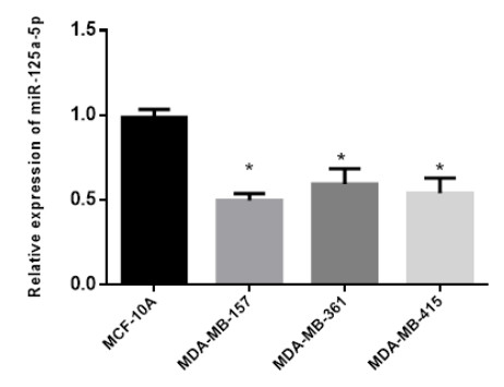
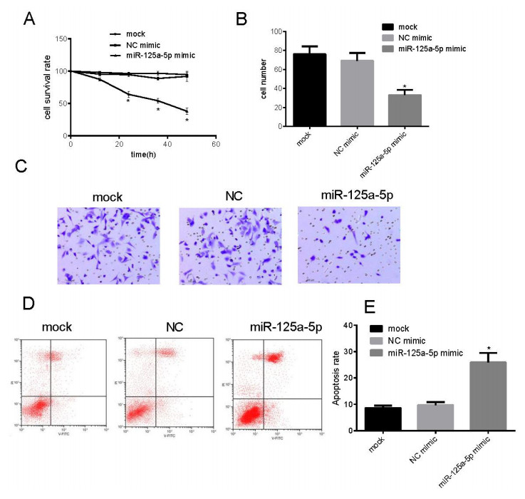
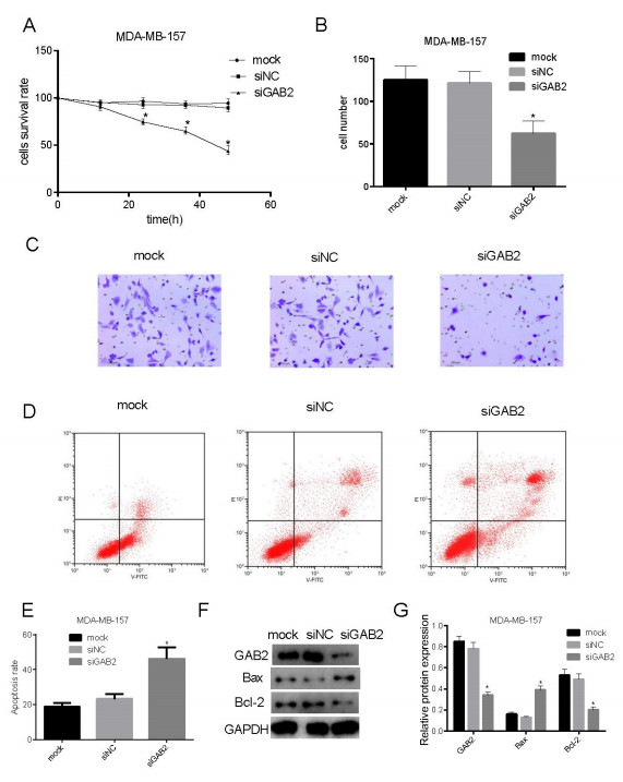
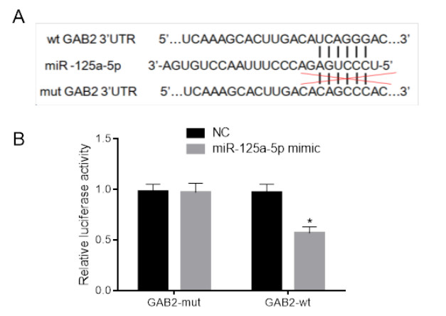
    Citation: Li-Bing Wang, Liang Feng, Jing He, Bo Liu, Jian-Guang Sun. MiR-125a-5p inhibits the proliferation and invasion of breast cancer cells and induces apoptosis by targeting GAB2[J]. Mathematical Biosciences and Engineering, 2019, 16(6): 6923-6933. doi: 10.3934/mbe.2019347

    Related Papers:

    [1] Shequn Gu, Jihui Luo, Wenxiu Yao . The regulation of miR-139-5p on the biological characteristics of breast cancer cells by targeting COL11A1. Mathematical Biosciences and Engineering, 2020, 17(2): 1428-1441. doi: 10.3934/mbe.2020073
    [2] Jun Xu, Bei Wang, Zhengtao Liu, Mingchun Lai, Mangli Zhang, Shusen Zheng . miR-223-3p regulating the occurrence and development of liver cancer cells by targeting FAT1 gene. Mathematical Biosciences and Engineering, 2020, 17(2): 1534-1547. doi: 10.3934/mbe.2020079
    [3] Wei Huo, Xiao-Min Zhu, Xin-Yan Pan, Min Du, Zhuo Sun, Zhi-Min Li . MicroRNA-527 inhibits TGF-β/SMAD induced epithelial-mesenchymal transition via downregulating SULF2 expression in non-small-cell lung cancer. Mathematical Biosciences and Engineering, 2019, 16(5): 4607-4621. doi: 10.3934/mbe.2019231
    [4] Xudan Ma, Qijun Zhang, Haihong Zhu, Kefeng Huang, Weina Pang, Qin Zhang . Establishment and analysis of the lncRNA-miRNA-mRNA network based on competitive endogenous RNA identifies functional genes in heart failure. Mathematical Biosciences and Engineering, 2021, 18(4): 4011-4026. doi: 10.3934/mbe.2021201
    [5] Guobing Lin, Cunming Zhang, Xuanyu Chen, Junwei Wang, Song Chen, Siyuan Tang, Tianqiang Yu . Identification of circulating miRNAs as novel prognostic biomarkers for bladder cancer. Mathematical Biosciences and Engineering, 2020, 17(1): 834-844. doi: 10.3934/mbe.2020044
    [6] Dongchen Lu, Wei Han, Kai Lu . Identification of key microRNAs involved in tumorigenesis and prognostic microRNAs in breast cancer. Mathematical Biosciences and Engineering, 2020, 17(4): 2923-2935. doi: 10.3934/mbe.2020164
    [7] Ming Zhang, Yingying Zhou, Yanli Zhang . High Expression of TLR2 in the serum of patients with tuberculosis and lung cancer, and can promote the progression of lung cancer. Mathematical Biosciences and Engineering, 2020, 17(3): 1959-1972. doi: 10.3934/mbe.2020104
    [8] Xu Guo, Yuanming Jing, Haizhou Lou, Qiaonv Lou . Effect and mechanism of long non-coding RNA ZEB2-AS1 in the occurrence and development of colon cancer. Mathematical Biosciences and Engineering, 2019, 16(6): 8109-8120. doi: 10.3934/mbe.2019408
    [9] Guohai Wang, Zhiyuan Hu . Icotinib inhibits proliferation and epithelial-mesenchymal transition of non-small cell lung cancer A549 cells. Mathematical Biosciences and Engineering, 2019, 16(6): 7707-7718. doi: 10.3934/mbe.2019386
    [10] Elena Izquierdo-Kulich, Margarita Amigó de Quesada, Carlos Manuel Pérez-Amor, Magda Lopes Texeira, José Manuel Nieto-Villar . The dynamics of tumor growth and cells pattern morphology. Mathematical Biosciences and Engineering, 2009, 6(3): 547-559. doi: 10.3934/mbe.2009.6.547
  • To investigate whether miR-125a-5p can inhibit the proliferation and invasion of breast cancer cells and induce apoptosis by targeting GAB2. Methods: qRT-PCR was used to detect the expression of miR-125a-5p in normal mammary epithelial cells and breast cancer cell lines; The miR-125a-5p overexpression plasmid was transiently transfected into MDA-MB-157 cells, and the proliferation, invasion and apoptosis of breast cancer cells were detected by CCK8 kit, Transwell chamber and flow cytometry, respectively; Gene silencing was used to knock down GAB2 gene in MDA-MB-157 cells, and the changes of proliferation, invasion, apoptosis and apoptosis-related proteins in breast cancer cells were detected by CCK8 kit, Transwell chamber, flow cytometry and western blot, respectively; The direct interaction between miR-125a-5p and GAB2 was detected by dual-luciferase reporter assay. The miR-125a-5p overexpression plasmid was transiently transfected into MDA-MB-157 cells, and the expression levels of GAB2 and apoptosis-related proteins were detected by western blot. Results: The expression of miR-125a-5p in breast cancer cell lines, MDA-MB-157 cells, MDA-MB-361 cells and MDA-MB-415 cells, was significantly lower than that in normal breast epithelial cells, MCF-10A cells; The proliferation and invasion ability of MDA-MB-157 cells transfected with miR-125a-5p were significantly inhibited, and the apoptosis rate was significantly increased; Since GAB2 knocked down, the proliferation and invasion ability of MDA-MB-157 cells were significantly inhibited, while the apoptosis rate was significantly increased, the Bax protein expression was significantly down-regulated, and the Bcl-2 protein expression was significantly up-regulated; The dual-luciferase reporter assay demonstrated that miR-125a-5p can specifically target GAB2. Transfected with miR-125a-5p, the GAB2 protein expression and Bax protein expression were significantly down-regulated, but the Bcl-2 protein expression was significantly up-regulated. Conclusion: miR-125a-5p inhibits the proliferation and invasion of breast cancer cells and induces their apoptosis by negatively regulating GAB2.


    Because of the high incidence and mortality, cancer has become the main cause of death, and has gradually developed as the primary health issue [1,2]. The incidence of breast cancer among women shows an increasing tendency year by year, ranking first in both developed and developing countries. In recent years, with the research of the invasion and metastasis mechanism of breast cancer by scholars, molecular targeted therapy has drawn extensive attention [3,4,5].

    MiRNAs are involved in the regulation of multiple signaling pathways, and play an important role in angiogenesis of tumors. Previous studies have shown that the expression of miR-125a-5p is abnormal in human tumors, such as non-small cell lung cancer [6], breast cancer [7], and gastric cancer [8]. Moreover, it was found that the regulatory pathway of HOXA11-AS/miR-125a-5p/Rab3D would promote OS metastasis [9]; miR-125a-5p could also serve as a potential therapeutic biomarker for CRC patients due to its role in inhibiting CRC cell migration, invasion and EMT [10]; Recently, Wang S and his team found that miR-125a-5p could inhibit the proliferation and EMT of gastric cancer by targeting FOXS1 [11]. On the whole, we assume that miR-125a-5p could play a similar role as tumor inhibitor by regulating related gene proteins.

    As a member of the Grb-associated binders (Gabs) family, GAB2 scaffold protein can activate downstream signal transduction pathway and play an important role in the proliferation, differentiation, invasion and metastasis of cancer cells after phosphorylation by tyrosine kinase. During the process of invasion and metastasis of breast cancer cells, GAB2 might be involved in related signal transduction [12,13]. It has been reported that miR-125a-5p can inhibit the invasion and metastasis of glioma cells and breast cancer cells by targeted regulating GAB2 [14,15], but whether miR-125a-5p can affect the proliferation and apoptosis of breast cancer cells by regulating GAB2 has not been reported. In this study, we investigated the molecular mechanism of miR-125a-5p in the migration, proliferation and apoptosis of breast cancer cells by altering the expression of miR-125a-5p in breast cancer cell line MDA-MB-157, so as to provide a new therapeutic target for clinical prevention and treatment of breast cancer cells.

    Human breast cancer cell strains, MDA-MB-157, MDA-MB-361 and MDA-MB-415, were purchased from Nanjing Cobioer Biotechnology Co., Ltd; RPMI-1640, fetal bovine serum and Annexin V-FITC/PI cell apoptosis detection kit were purchased from Beijing Solarbio Science & Technology Co., Ltd; MEM medium was purchased from Gibco; Bax, Bcl-2, GAB2 and GADPH antibodies were purchased from Santa Cruz company; miR-125a-5p overexpression Plasmids and gene vectors of dual-luciferase reporter were constructed by Obio Technology Corp., Ltd in Shanghai, China; LipofectamineTM2000 and Trizol lysate were purchased from Invitrogen company; Promega Dual-Luciferase system detection kit was purchased from Hang Seng Biotechnology Co., Ltd; siRNA plasmids of GAB2 were purchased from Shanghai GenePharma; Transwell chamber was purchased from Milipore; the detection reagent for CCK8 proliferation was purchased from DOJINDO.

    MDA-MB-157 cells, MDA-MB-361 cells and MDA-MB-415 cells were cultured in DMEM medium containing 10% FBS, DMEM medium containing 10% FBS and L-15 medium containing 10% FBS, respectively. Human normal breast epithelial cells, MCF-10A cells, were cultured in DMEM/F12 (1:1) medium containing 10% FBS. Following the instruction of LipofectamineTM2000 transfection reagent, the cells were transfected. The experimental groups were as below: 1) mock group, blank control group with only transfection reagent; 2) NC mimic group, negative control group for transfection of independent sequence; 3) miR-125a-5p mimic group, the experimental group for transfection of miR-125a-5p plasmids.

    Cells in each group were inoculated into six-well plate with a density of 3 × 10⁵. When the growth density of cells reached 90%, we diluted them respectively and added 100 nmol/L plasmids to co-transfect with LipofectamineTM2000. After 5 minutes at room temperature, the cells were mixed and dripped into the cell culture pore after 20 minutes. Shake the plate gently to make the cells mixed, and continuous culture them in a culture box with 5% CO2 at 37℃. After 6 hours, the culture medium was replaced and the cells were transfected for 48 hours and collected for subsequent experiments.

    MDA-MB-157 cells in logarithmic phase were selected and divided into the following experimental groups: 1) mock group, blank control group with only transfection reagents; 2) NC group, the experimental group for transfection of Scr siRNA plasmids; 3) siGAB2 group, the experimental group for transfection of siGAB2 plasmids. Dilute 20μΜ siRNA of 8μL and incubate it at room temperature for 5 minutes after gently mixed with 250μL serum-free DMEM. Then mix the siRNA diluent with LipofectamineTM2000 with a total volume of 500 mL, incubated at room temperature for 20 minutes. The medium was replaced before transfection, and the corresponding reagents were added according to the experimental groups. The transfection time was 48 hours.

    With LipofectamineTM2000 transfection reagent, wild GAB2 (wt-GAB2) and mutant GAB2 (mut-GAB2) were co-transfected into MDA-MB-157 cells with blank plasmids and over-expressed miR-125a-5p plasmids, respectively. After 48 hours of incubation, the cells were collected and dual-luciferase was detected with Promega Dual-Luciferase system detection kit, which was performed as previously described [16]. Precipitation was washed by PBS twice, and lysed with cell lysate for 15 minutes, then it was centrifuged and the supernatant was taken. LAR II solution was added to rapidly determine the fluorescence intensity of samples. Finally, the reaction termination solution was added to detect the ranilla luciferase reaction intensity.

    Real-time Fluorescence Quantitative PCR analysis was performed as previously described [17]. Trizol was used to lyse breast cancer cells and normal breast epithelial cells. Extract total RNA and determine its purity and concentration. The reverse transcription was carried out by adding the primer miR-125a-5p. U6 was selected as internal reference for qRT-PCR. The procedure was as below: 94℃ for 4 minutes, 94℃ for 20 seconds, 60℃ for 30 seconds, 72℃ for 30 seconds. Repeat this procedure 35 times.

    miR-125a-5p

    Forward Primer: 5'-ACACTCCAGCTGGGTCCCTGAGACCCTTTAA-3';

    Reverse Primer: 5'-CTCAACTGGTGTCGTGGAGT-3'.

    U6

    Forward Primer: 5'-CTCGC TTCGGCAGCACA-3';

    Reverse Primer: 5'-AACGCTTCACGAATTTGCGT-3'.

    Based on the above experimental grouping and transfection methods, total protein was extracted after lysis, and conducted electrophoresis with 12% SDS-PAGE gel, then membrane transferred at 300mA for two hours. Primary antibodies, GAB2 (1:1000), Bax (1:1000), bcl-2 (1:2000), GADPH (1:1000), were added after sealed and then incubated at 4℃ overnight. After washing membrane with TBST, second antibodies were added and incubated at room temperature for one hour. Then wash again and add ECL. The membrane was Exposed with X-ray film, developed with developer, fixed with fixation in dark room, then rinse and dry it, scan image, and repeat experiment independently for 3 times [18].

    Based on the above experimental grouping and transfection methods, the cells were inoculated into the upper chamber of 24-well Transwell chamber at the density of 2*105 cell/well. Matrigel glue was laid on the upper chamber and the culture medium with a volume of 600μL containing 10% FBS was added to the lower chamber. After 24 hours of incubation, carefully remove the Transwell chamber and fix it for 20 minutes with 4% polyformaldehyde solution. Then wash it with PBS solution, and wipe out the cells on the surface of the chamber with cotton swabs to crystallize. After purple staining, microscopic observation was carried out. Select six visual fields of each group to photograph and count, and calculate an average. The experiment was repeated independently for three times [19].

    Based on the above experimental grouping and transfection methods, the cell proliferation was detected by CCK8 method. The cells were inoculated into 96-well plate at the density of 8000 cells/well. The method of experimental group dividing was the same as before. After 12, 24, 36 and 48 hours of transfection, the plate was replaced with a medium containing 10% CCK8, respectively. After 4 hours of continuous culture in the incubator, detect the OD value at 450 nm with enzyme labeling instrument, calculate the increment rate and draw the cell proliferation curve. Cell proliferation rate = (OD value of transfected cells/OD value of the blank control) * 100%.

    Apoptosis rate analysis was performed as previously described [20]. Based on the above experimental grouping and transfection methods, Annexin V-FITC/PI cell apoptosis detection kit was used to detect the apoptosis rate following the instruction. The method of experimental group dividing was the same as before. After 48 hours of transfection, the cells were collected and precipitated with 100μL 1 × Binding Buffer, then incubated with 5μL Annexin V-FITC solution at room temperature away from light for 10 minutes, and then incubated with 5μL PI solution in the same environment for 5 minutes. Finally, PBS solution was added to a total volume of 500μL. Gently mix and use Accuri C6 flow cytometry to detect the apoptosis rate of cells in each group.

    Relevant data was analyzed statistically by using SPSS21.0, and tested with independent sample t. P < 0.05 was considered to have significant differences with statistical significance.

    In order to investigate the expression of miR-125a-5p in normal breast epithelial cells and breast cancer cell lines, we used qRT-PCR to detect the expression of miR-125a-5p in each group. Results as shown in Figure 1. The expression of miR-125a-5p in breast cancer cell lines, MDA-MB-157, MDA-MB-361 and MDA-MB-415, was significantly lower than that in normal breast epithelial cells, MCF-10A (P < 0.05). Therefore, MDA-MB-157 cell strain was used for subsequent experiments.

    Figure 1.  Expression levels of miR-125a-5p in MCF-10A, MDA-MB-157, MDA-MB-361 and MDA-MB-415 cells (x ± s, n = 3). *P < 0.05 vs MCF-10A.

    CCK8 kit was used to detect the effect of miR-125a-5p on the proliferation of MDA-MB-157 cells. As shown in Figure 2A, the proliferation rate of MDA-MB-157 cells in the miR-125a-5p mimic group was significantly lower than that in the mock group and NC mimic group, and the difference had statistical significance (P < 0.05). Transwell assay was used to detect the effect of miR-125a-5p on the invasion of MDA-MB-157 cells. As shown in Figure 2B, the number of MDA-MB-157 cells invading in the miR-125a-5p mimic group was significantly lower than that in the mock group and NC mimic group, and the difference had statistical significance (P < 0.05). Flow cytometry was used to detect the effect of miR-125a-5p on the apoptosis of MDA-MB-157 cells. As shown in Figure 2C and D, the apoptotic number of MDA-MB-157 cells in miR-125a-5p mimic group was significantly higher than that in mock group and NC mimic group, and the difference had statistical significance (P < 0.05). The results showed that promoting the expression of miR-125a-5p could inhibit the proliferation, invasion and apoptosis of MDA-MB-157 cells.

    Figure 2.  miR-125a-5p inhibits the proliferation and invasion of breast cancer cells and induces their apoptosis (x ± s, n = 3). A: The proliferation of MDA-MB-157 cell transfected with mock、NC mimic and miR-125a-5p mimic for 12h, 24h, 36h and 48h were detected by CCK8 kit; B, C: The Invasion of MDA-MB-157 cell transfected with mock、NC mimic and miR-125a-5p mimic were detected by Transwell invasion assay; D, E: The apoptosis of MDA-MB-157 cell transfected with mock、NC mimic and miR-125a-5p mimic were detected flow cytometry. *P < 0.05 vs MDA-MB-157/mock or MDA-MB-157/NC.

    CCK8 kit was used to detect the effect of GAB2 on the proliferation of MDA-MB-157 cells. As shown in Figure 3A, the proliferation rate of MDA-MB-157 cells in siGAB2 group was significantly lower than that in mock group and siNC group, and the difference had statistical significance (P < 0.05). Transwell assay was used to detect the effect of silencing GAB2 on the invasion of MDA-MB-157 cells. As shown in Figure 3B and C, the number of MDA-MB-157 cells invading in siGAB2 group was significantly lower than that in mock group and siNC group, and the difference had statistical significance (P < 0.05). Flow cytometry was used to detect the effect of GAB2 on the apoptosis of MDA-MB-157 cells. As shown in Figure 3D and E, the apoptotic number of MDA-MB-157 cells in GAB2 group was significantly higher than that in mock group and siNC group, and the difference had statistical significance (P < 0.05). The expression of apoptosis-associated proteins, Bax and Bcl-2, was detected by Western blot. As shown in Figure 3F, compared with mock group and siNC group, the expression of Bax protein in GAB2 group was significantly down-regulated, and the expression of Bcl-2 protein was significantly up-regulated (P < 0.05). These results suggested that the expression of silencing GAB2 could inhibit the proliferation, invasion and apoptosis of MDA-MB-157 cells.

    Figure 3.  Knockdown of GAB2 inhibits the proliferation and invasion of breast cancer cells and induces their apoptosis (x ± s, n = 3). A: The proliferation of MDA-MB-157 cell transfected with mock、siNC and siGAB2 for 12h, 24h, 36h and 48h were detected by CCK8 kit; B, C: The Invasion of MDA-MB-157 cell transfected with mock、siNC and siGAB2 were detected by Transwell invasion assay; D, E: The apoptosis of MDA-MB-157 cell transfected with mock、siNC and siGAB2 were detected flow cytometry; F, G: The expression of Bax and Bcl-2 of MDA-MB-157 cell transfected with mock、siNC and siGAB2 were detected by western blot. *P < 0.05 vs mock or siNC.

    According to the Targetscan.org website, we could predict the regional sequence where miR-125a-5p could be directly targeted combined with GAB2 3'-UTR, as shown in Figure 4A below. The results of dual-luciferase assay showed in Figure 4B indicated that the fluorescence intensity of co-transfection group with GAB2-wt and miR-125a-5p mimic was significantly lower than that with GAB2-mut and miR-125a-5p mimic, and GAB2-wt and NC mimic (P < 0.05). The results showed that miR-125a-5p could be targeted inhibit the expression of GAB2.

    Figure 4.  miR-125a-5p down-regulate the expression of GAB2、Bcl-2 and up-regulate the expression of Bax. A: The binding sequence of miR-125a-5p and the sequence of targeted 3'-UTR of GAB2; B: The result of dual luciferase reporter assay about the interaction between miR-125a-5p and GAB2.

    Breast cancer is one of the most common multiple malignant tumors in women worldwide, which seriously threatens human health and quality of life. Statistically, both the incidence and mortality of breast cancer in China have been increasing year by year in recent years. With the continuous development of medical level, the detection rate of breast cancer has been significantly improved. However, most patients have had lymph node metastasis at the time of detection, so the treatment effect is not optimistic and inhibiting the metastasis of breast cancer cells has become the key to improve the survival rate of patients [21]. Invasion and metastasis are considered to be main reasons for breast cancer recurrence and deaths due to breast cancer. Therefore, the research on the molecular mechanism of breast cancer is still a hot topic in recent years.

    GAB2 is a potential oncogene, and its abnormal amplification is closely related to the occurrence and metastasis of breast cancer. Studies have shown that GAB2 is overexpressed in lung cancer, ovarian cancer, chronic myelogenous leukemia, multiple primary breast cancer and various breast cancer cell lines [22,23,24]. In this study, we found that knocking down GAB2 expression by RNAi technology could inhibit the invasion and proliferation of breast cancer cells. Therefore, GAB2 can be used as a therapeutic target to inhibit the invasion and proliferation of breast cancer, and reduce the expression of GAB2 in cancer cells, so as to inhibit the invasion and proliferation of breast cancer cells.

    MiRNA is a class of single-stranded non-coding RNA with about 20 bases. Many studies have shown that miRNA can inhibit or promote the invasion and proliferation of tumors by regulating the expression of related target genes in organisms [25]. At present, miRNA have become a new potential tumor marker. Therefore, exploring the role of miRNA in the molecular mechanism of breast cancer has far-reaching significance for the prevention and treatment of clinical breast cancer. miR-125a-5p is a member of the miR-125 family, which originates from the 5'end of pre-miR-125a. More and more studies have shown that mature miR-125a-5p could inhibit the invasion and metastasis of many malignant tumors, such as liver cancer, colon cancer, breast cancer and non-small cell lung cancer, playing the role of tumor suppressor [26,27,28].

    Through this experiment, we can find that miR-125a-5p could inhibit the proliferation and invasion of breast cancer cells by negatively regulating GAB2, and induce the apoptosis of breast cancer cells by regulating the expression of Bax and Bcl-2 proteins in apoptotic pathway.

    The authors declare that they have no conflict of interest.


    [1] W. Chen, R. Zheng, P. D. Baade, et al., Cancer statistics in China, 2015, CA-Cancer J. Clin., 66 (2016), 115–132.
    [2] R. L. Siegel, K. D. Miller and A. Jemal, Cancer Statistics, 2017, CA-Cancer J. Clin., 67 (2017), 7–30.
    [3] E. Dirican and M. Akkiprik, Phosphatidylinositol 3-kinase regulatory subunit 1 and phosphatase and tensin homolog as therapeutic targets in breast cancer, Tumour Biol., 39 (2017), 1010428317695529.
    [4] S. Nagini, Breast cancer: Current molecular therapeutic targets and new players, Anticancer Agents Med. Chem., 17 (2017), 152–163.
    [5] M. Untch, G. E. Konecny, S. Paepke, et al., Current and future role of neoadjuvant therapy for breast cancer, Breast, 23 (2014), 526–537.
    [6] R. J. Wang, Y. H. Zheng, P. Wang, et al., Serum miR-125a-5p, miR-145 and miR-146a as diagnostic biomarkers in non-small cell lung cancer, Int. J. Clin. Exp. Pathol., 8 (2015), 765–771.
    [7] H. He, F. Xu, W. Huang, et al., miR-125a-5p expression is associated with the age of breast cancer patients, Genet. Mol. Res., 14 (2015), 17927–17933.
    [8] Y. Xu, Z. Huang and Y. Liu, Reduced miR-125a-5p expression is associated with gastric carcinogenesis through the targeting of E2F3, Mol. Med. Rep., 10 (2014), 2601–2608.
    [9] K. Cao, Y. Fang, H. Wang, et al., The lncRNA HOXA11-AS regulates Rab3D expression by sponging miR-125a-5p promoting metastasis of osteosarcoma, Cancer Manag. Res., 11 (2019), 4505–4518.
    [10] L. Tang, L. Zhou, S. Wu, et al., miR-125a-5p inhibits colorectal cancer cell epithelial-mesenchymal transition, invasion and migration by targeting TAZ, Oncotargets Ther., 12 (2019), 3481–3489.
    [11] S. Wang, L. Ran, W. Zhang, et al., FOXS1 is regulated by GLI1 and miR-125a-5p and promotes cell proliferation and EMT in gastric cancer, Sci. Rep., 9 (2019), 5281.
    [12] Y. Chen, Q. Liu, M. Wu, et al., GAB2 promotes cell proliferation by activating the ERK signaling pathway in hepatocellular carcinoma, Tumour Biol., 37 (2016), 11763–11773.
    [13] C. B. Ding, W. N. Yu, J. H. Feng, et al., Structure and function of Gab2 and its role in cancer (Review), Mol. Med. Rep., 12 (2015), 4007–4014.
    [14] L. Sun, Y. Liu, X. Li, et al., Gab2-Akt-ARK5 signaling pathway is associated with the invasion of glioma cells, Chin. J. Clin. Onco., 41 (2014), 551–554.
    [15] X. W. Xu, P. R. Li, Z. S. Zhang, et al., miR-125a-5p inhibits the migration of breast cancer cells by targeting GAB2, Chin. J. Biochem. Mol. Biol., (2017).
    [16] X. J. Li, Z. F. Li, Y. Y. Xu, et al., microRNA-374 inhibits proliferation and promotes apoptosis of mouse melanoma cells by inactivating the Wnt signalling pathway through its effect on tyrosinase, J. Cell. Mol. Med., (2019).
    [17] T. Matsumura, K. Sugimachi, Y. Takahashi, et al., Clinical significance of GAB2, a scaffolding/docking protein acting downstream of EGFR in human colorectal cancer, Ann. Surg. Oncol., 21 Suppl 4 (2014), S743–749.
    [18] N. Leblanc, J. Harquail, N. Crapoulet, et al., Pax-5 inhibits breast cancer proliferation through MiR-215 up-regulation, Anticancer Res., 38 (2018), 5013–5026.
    [19] Y. Yang, D. P. Li, N. Shen, et al., TPX2 promotes migration and invasion of human breast cancer cells, Asian Pac. J. Trop. Med., 8 (2015), 1064–1070.
    [20] M. Frohlich, N. Schafer, M. Caspers, et al., Temporal phenotyping of circulating microparticles after trauma: a prospective cohort study, Scand. J. Trauma. Resusc. Emerg. Med., 26 (2018), 33.
    [21] C. Cedolini, S. Bertozzi, A. P. Londero, et al., Type of breast cancer diagnosis, screening, and survival, Clin. Breast Cancer, 14 (2014), 235–240.
    [22] S. J. Davis, K. E. Sheppard, M. S. Anglesio, et al., Enhanced GAB2 expression is associated with improved survival in high-grade serous ovarian cancer and sensitivity to PI3K inhibition, Mol. Cancer Ther., 14 (2015), 1495–1503.
    [23] S. Halbach, K. T. Rigbolt, F. U. Wohrle, et al., Alterations of Gab2 signalling complexes in imatinib and dasatinib treated chronic myeloid leukaemia cells, Cell. Commun. Signal, 11 (2013), 30.
    [24] X. L. Xu, X. Wang, Z. L. Chen, et al., Overexpression of Grb2-associated binder 2 in human lung cancer, Int. J. Biol. Sci., 7 (2011), 496–504.
    [25] D. Park, S. C. Lee, J. W. Park, et al., Overexpression of miR-17 in gastric cancer is correlated with proliferation-associated oncogene amplification, Pathol. Int., 64 (2014), 309–314.
    [26] Z. Cai, J. Li, Q. Zhuang, et al., MiR-125a-5p ameliorates monocrotaline-induced pulmonary arterial hypertension by targeting the TGF-β1 and IL-6/STAT3 signaling pathways, Exp. Mol. Med., 50 (2018), 45.
    [27] D. W. Melton, X. Lei, J. A. Gelfond, et al., Dynamic macrophage polarization-specific miRNA patterns reveal increased soluble VEGF receptor 1 by miR-125a-5p inhibition, Physiol. Genomics, 48 (2016), 345–360.
    [28] K. Odar, E. Bostjancic, N. Gale, et al., Differential expression of microRNAs miR-21, miR-31, miR-203, miR-125a-5p and miR-125b and proteins PTEN and p63 in verrucous carcinoma of the head and neck, Histopathology, 61 (2012), 257–265.
  • This article has been cited by:

    1. Wanqing Weng, Zhongjing Zhang, Weiguo Huang, Xiangxiang Xu, Boda Wu, Tingbo Ye, Yunfeng Shan, Keqing Shi, Zhuo Lin, Identification of a competing endogenous RNA network associated with prognosis of pancreatic adenocarcinoma, 2020, 20, 1475-2867, 10.1186/s12935-020-01243-6
    2. Mimi Xu, Jiandong Zhan, Jiaxuan Xie, Lijun Zhu, Liangsi Chen, Xiaoning Luo, Xiaoli Sheng, Tao Liu, Siyi Zhang, Zhongming Lu, MiR-125a-5p inhibits cell proliferation, cell cycle progression, and migration while promoting apoptosis in head and neck cancers by targeting ERBB3, 2020, 03858146, 10.1016/j.anl.2020.10.001
    3. Lantang Huang, Ruimin Liu, Peiyi Zhou, Yingpu Tian, Zhongxian Lu, miR-9 and miR-181a Target Gab2 to Inhibit the Proliferation and Migration of Hepatocellular Carcinoma HepG2 Cells, 2022, 13, 2073-4425, 2152, 10.3390/genes13112152
    4. Dongmei Li, Limei Chen, Xiaofang Zhang, Yanhua Wang, Chuansheng Huang, Jianglong Li, Feilong He, Wenxing He, miR‑125a‑5p reverses epithelial‑mesenchymal transition and restores drug sensitivity by negatively regulating TAFAZZIN signaling in breast cancer, 2021, 24, 1791-2997, 10.3892/mmr.2021.12452
    5. Yongjun Ren, Caixia Zhao, Yi He, Xuli Min, Hao Xu, Xiao Hu, RPARP-AS1/miR125a-5p Axis Promotes Cell Proliferation, Migration and Invasion in Colon Cancer, 2021, Volume 14, 1178-6930, 5035, 10.2147/OTT.S304494
    6. Kang Gao, Tao Zhou, YingChun Yin, XiaoJie Sun, HePing Jiang, TangYue Li, Atorvastatin inhibits glioma glycolysis and immune escape by modulating the miR-125a-5p/TXLNA axis, 2024, 161, 1601-5223, 10.1186/s41065-024-00349-5
    7. Shih‐Wen Kao, Wei‐Wen Kuo, Bruce Chi‐Kang Tsai, Ebro Chawesa Mwakalinga, Thomas Gabriel Mhone, Ming‐Cheng Chen, Chia‐Hua Kuo, Yi‐Sheng Liu, Shinn‐Zong Lin, Chih‐Yang Huang, Hepatocellular Carcinoma Cells in Humans Exhibit Resistance to Suberoylanilide Hydroxamic Acid (SAHA) Owing to the Diminished Level of Hsa‐miR‐125a‐5p, 2025, 105, 1747-0277, 10.1111/cbdd.70040
    8. Yuliya A. Veryaskina, Sergei E. Titov, Igor B. Kovynev, Sofya S. Fyodorova, Olga V. Berezina, Igor P. Zhurakovskij, Oksana V. Antonenko, Sergei A. Demakov, Pavel S. Demenkov, Pavel S. Ruzankin, Anton S. Tarasenko, Tatiana I. Pospelova, Igor F. Zhimulev, MicroRNAs in Diffuse Large B-Cell Lymphoma (DLBCL): Biomarkers with Prognostic Potential, 2025, 17, 2072-6694, 1300, 10.3390/cancers17081300
  • Reader Comments
  • © 2019 the Author(s), licensee AIMS Press. This is an open access article distributed under the terms of the Creative Commons Attribution License (http://creativecommons.org/licenses/by/4.0)
通讯作者: 陈斌, bchen63@163.com
  • 1. 

    沈阳化工大学材料科学与工程学院 沈阳 110142

  1. 本站搜索
  2. 百度学术搜索
  3. 万方数据库搜索
  4. CNKI搜索

Metrics

Article views(5899) PDF downloads(533) Cited by(8)

Figures and Tables

Figures(4)

/

DownLoad:  Full-Size Img  PowerPoint
Return
Return

Catalog