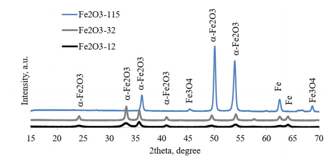
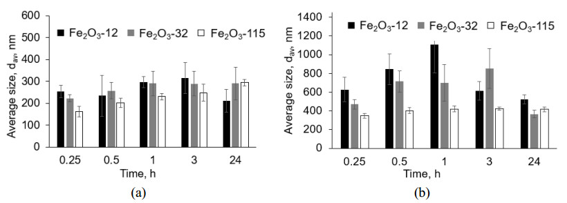
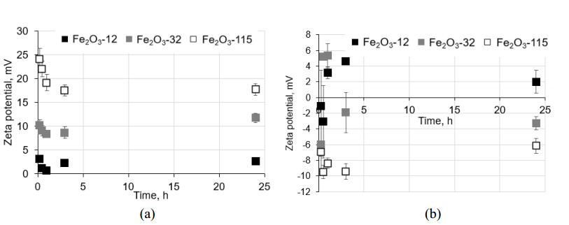
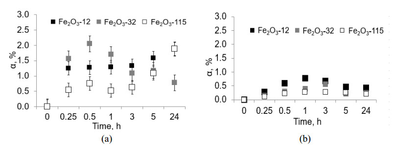
Despite high medical and biological potential, the penetration of iron oxide nanoparticles (NPs) into a human body can cause their dissolution with subsequent accumulation of highly toxic iron compounds. The paper describes the agglomeration and dissolution behavior of differently sized α-Fe2O3 NPs in the simplest biological solutions. The average sizes of the initial NPs according to the BET analysis are 12, 32, and 115 nm. Within 30–60 min exposure, the particle size and concentration of iron released into the solutions increases in the suspensions, accompanied by an intensive change of NPs surface charge. After an hour of exposure, the colloidal properties do not change significantly, although the dissolution degree ambiguously fluctuates. It has been shown that the agglomeration of the particles in the simplest pulmonary fluid is lower than in the simplest sweat fluid, compared to the dissolution degree, which is much higher in the pulmonary fluid than in the sweat. The colloidal stability of suspensions reduces with a decrease of NPs' size, e.g., the average size of particles is 315,289, and 248 nm, while zeta potential is 2, 9, and 17 mV, respectively for 12, 32, and 115 nm NPs in 3-hour suspensions. It has been found that 24 h dissolution degree of α-Fe2O3 NPs reaches 2.3% and 0.4%, respectively, in the simplest pulmonary and sweat fluids. The mechanism of dissolution of hematite NPs in the slightly acidic and acidic mediums is proposed.
Citation: Anna Godymchuk, Alexey Ilyashenko, Yury Konyukhov, Peter Ogbuna Offor, Galiya Baisalova. Agglomeration and dissolution of iron oxide nanoparticles in simplest biological media[J]. AIMS Materials Science, 2022, 9(4): 642-652. doi: 10.3934/matersci.2022039
Despite high medical and biological potential, the penetration of iron oxide nanoparticles (NPs) into a human body can cause their dissolution with subsequent accumulation of highly toxic iron compounds. The paper describes the agglomeration and dissolution behavior of differently sized α-Fe2O3 NPs in the simplest biological solutions. The average sizes of the initial NPs according to the BET analysis are 12, 32, and 115 nm. Within 30–60 min exposure, the particle size and concentration of iron released into the solutions increases in the suspensions, accompanied by an intensive change of NPs surface charge. After an hour of exposure, the colloidal properties do not change significantly, although the dissolution degree ambiguously fluctuates. It has been shown that the agglomeration of the particles in the simplest pulmonary fluid is lower than in the simplest sweat fluid, compared to the dissolution degree, which is much higher in the pulmonary fluid than in the sweat. The colloidal stability of suspensions reduces with a decrease of NPs' size, e.g., the average size of particles is 315,289, and 248 nm, while zeta potential is 2, 9, and 17 mV, respectively for 12, 32, and 115 nm NPs in 3-hour suspensions. It has been found that 24 h dissolution degree of α-Fe2O3 NPs reaches 2.3% and 0.4%, respectively, in the simplest pulmonary and sweat fluids. The mechanism of dissolution of hematite NPs in the slightly acidic and acidic mediums is proposed.

| [1] |
Dadfar SM, Roemhild K, Drude NI, et al. (2019) Iron oxide nanoparticles: Diagnostic, therapeutic and theranostic applications. Adv Drug Delivery Rev 138: 302–325. https://doi.org/10.1016/j.addr.2019.01.005 doi: 10.1016/j.addr.2019.01.005

|
| [2] |
Laurent S, Forge D, Port M (2008) Magnetic iron oxide nanoparticles: synthesis, stabilization, vectorization, physicochemical characterizations, and biological applications. Chem Rev 108: 2064–2110. https://doi.org/10.1021/cr068445e doi: 10.1021/cr068445e
|
| [3] |
Arias LS, Pessan JP, Vieira APM, et al. (2018) Monteiro Iron oxide nanoparticles for biomedical applications: a perspective on synthesis, drugs, antimicrobial activity, and toxicity. Antibiotics (Basel) 7: 46. https://doi.org/10.3390/antibiotics7020046 doi: 10.3390/antibiotics7020046
|
| [4] |
Attarad A, Hira Z, Muhammad Z (2016) Synthesis, characterization, applications, and challenges of iron oxide nanoparticles. Nanotechnol Sci Appl 9: 49–67. https://doi.org/10.2147/NSA.S99986 doi: 10.2147/NSA.S99986
|
| [5] |
Roelofs F, Vogelsberger W (2006) Dissolution kinetics of nanodispersed γ-alumina in aqueous solution at different pH: Unusual kinetic size effect and formation of a new phase. J Colloid Interf Sci 303: 450–459. https://doi.org/10.1016/j.jcis.2006.08.016 doi: 10.1016/j.jcis.2006.08.016
|
| [6] |
Baalousha M (2009) Aggregation and disaggregation of iron oxide nanoparticles: Influence of particle concentration, pH and natural organic matter. Sci Total Environ 407: 2093–2101. https://doi.org/10.1016/j.scitotenv.2008.11.022 doi: 10.1016/j.scitotenv.2008.11.022
|
| [7] |
Strehlau JH, Toner BM, Arnold WA, et al. (2017) Accessible reactive surface area and abiotic redox reactivity of iron oxyhydroxides in acidic brines. Geochim Cosmochim Ac 197: 345–355. https://doi.org/10.1016/j.gca.2016.10.015 doi: 10.1016/j.gca.2016.10.015
|
| [8] |
Liu J, Dai C, Hu Y (2018) Aqueous aggregation behavior of citric acid coated magnetite nanoparticles: Effects of pH, cations, anions, and humic acid. Environ Res 161: 49–60. https://doi.org/10.1016/j.envres.2017.10.045 doi: 10.1016/j.envres.2017.10.045
|
| [9] |
Maenosono S, Suzuki T, Saita S (2007) Mutagenicity of water-soluble FePt nanoparticles in Ames test. J Toxicol Sci 32: 575–579. https://doi.org/10.2131/jts.32.575 doi: 10.2131/jts.32.575
|
| [10] |
Waite TD, Morel FMM (1984) Photoreductive dissolution of colloidaliron oxide: Effect of citrate. J Colloid Interf Sci 102: 121–137. https://doi.org/10.1016/0021-9797(84)90206-6 doi: 10.1016/0021-9797(84)90206-6
|
| [11] |
Xu N, Gao Y (2008) Characterization of hematite dissolution affected by oxalate coating, kinetics and pH. Appl Geochem 23: 783–793. https://doi.org/10.1016/j.apgeochem.2007.12.026 doi: 10.1016/j.apgeochem.2007.12.026
|
| [12] |
Borer PM, Sulzberger B, Reichar P, et al. (2005) Effect of siderophores on the light-induced dissolution of colloidal iron(Ⅲ) (hydr)oxides. Mar Chem 93: 179–193. https://doi.org/10.1016/j.marchem.2004.08.006 doi: 10.1016/j.marchem.2004.08.006
|
| [13] |
Bligh MW, Waite TD (2011) Formation, reactivity, and aging of ferric oxide particles formed from Fe(Ⅱ) and Fe(Ⅲ) sources: Implications for iron bioavailability in the marine environment. Geochim Cosmochim Ac 75: 7741–7758. https://doi.org/10.1016/j.gca.2011.10.013 doi: 10.1016/j.gca.2011.10.013
|
| [14] |
Godymchuk A, Papina I, Karepina E, et al. (2019) Agglomeration of iron oxide nanoparticles: pH effect is stronger than amino acid acidity. J Nanopart Res 21: 208. https://doi.org/10.1007/s11051-019-4634-y doi: 10.1007/s11051-019-4634-y
|
| [15] |
Marques MRC, Loebenberg R, Almukainzi M (2011) Simulated biological fluids with possible application in dissolution testing. Dissolut Technol 18: 15–28. https://doi.org/10.14227/DT180311P15 doi: 10.14227/DT180311P15
|
| [16] |
Midander K, Julander A, Kettelarij J (2016) Testing in artificial sweat—Is less more? Comparison of metal release in two different artificial sweat solutions. Regul Toxicol Pharm 81: 381–386. https://doi.org/10.1016/j.yrtph.2016.09.021 doi: 10.1016/j.yrtph.2016.09.021
|
| [17] |
Langevin D, Raspaud E, Mariot S, et al. (2018) Towards reproducible measurement of nanoparticle size using dynamic light scattering: Important controls and considerations NanoImpact 10: 161–167. https://doi.org/10.1016/j.impact.2018.04.002 doi: 10.1016/j.impact.2018.04.002
|
| [18] |
Abzhanova D, Godymchuk A, Gusev A, et al. (2016) Exposure of nano- and ultrafine Ni particles to synthetic biological solutions: predicting fate-related dissolution and accumulation. EJNM 8: 203–212. https://doi.org/10.1515/ejnm-2016-0021 doi: 10.1515/ejnm-2016-0021
|
| [19] |
Zhang YC, Tang JY, Xiao YH (2008) Controllable synthesis and magnetic properties of pure hematite and maghemite nanocrystals from a molecular precursor. J Alloy Compd 462: 24–28. https://doi.org/10.1016/j.jallcom.2007.07.115 doi: 10.1016/j.jallcom.2007.07.115
|
| [20] |
Prakash R, Fanselau K, Mandal TK (2013) A facile synthesis of a carbon-encapsulated Fe3O4 nanocomposite and its performance as anode in lithium-ion batteries. Beilstein J Nanotech 4: 699–704. https://doi.org/10.3762/bjnano.4.79 doi: 10.3762/bjnano.4.79
|
| [21] |
Mushtaq D (2007) The synthesis of maghemite and hematite (γ-Fe2O3, α-Fe2O3) nanospheres. Mater Sci Forum 534: 157–160. https://doi.org/10.4028/www.scientific.net/MSF.534-536.157 doi: 10.4028/www.scientific.net/MSF.534-536.157
|
| [22] |
Lanzl CA, Baltrusaitis J, Cwiertny DM (2012) Dissolution of hematite nanoparticle aggregates: influence of primary particle size, dissolution mechanism, and solution pH Langmuir 28: 15797–15808. https://doi.org/10.1021/la3022497 doi: 10.1021/la3022497
|
| [23] |
Favela-Camacho SE, Perez-Robles JF, Garcıa-Casillas PE, et al. (2016) Stability of magnetite nanoparticles with different coatings in a simulated blood plasma J Nanopart Res 18: 176. https://doi.org/10.1007/s11051-016-3482-2 doi: 10.1007/s11051-016-3482-2
|
| [24] |
Vodyanitskii YN (2013) Dissolution of magnetite and redistribution of heavy metals in urban soils (model experiment). Eurasian Soil Sc 46: 672–680. https://doi.org/10.1134/S1064229313060112 doi: 10.1134/S1064229313060112
|
| [25] | Wagman DD, Evans WH, Parker VB, et al. (1989) The NBS tables of chemical thermodynamic properties. Selected values for inorganic and C1 and C2 organic substances in SI units. Wanshington, American Society of Chemistry. Available from: https://srd.nist.gov/JPCRD/jpcrdS2Vol11.pdf. |
| [26] |
Mostovshchikov AV, Ilyin AP, Azanov AA, et al. (2016) The energy stored in the aluminum nanopowder irradiated by electron beam. Key Eng Mater 685: 639–642. https://doi.org/10.4028/www.scientific.net/KEM.685.639 doi: 10.4028/www.scientific.net/KEM.685.639
|
| [27] | SenGupta AK (2017) Table of solubility product constants at 25 ℃, Ion Exchange in Environmental Processes: Fundamentals, Applications and Sustainable Technology, John Wiley & Sons. |
| [28] |
Kuzin AV, Gorichev IG, Lainer YA (2013) Stimulating effect of phosphate ions on the dissolution kinetics of iron oxides in an acidic medium. Russ Metall 2013: 652–657. https://doi.org/10.1134/S0036029513090073 doi: 10.1134/S0036029513090073
|
| [29] |
Kreyling W G, Semmler M, Erbe F (2002) Translocation of ultrafine insoluble iridium particles from lung epithelium to extrapulmonary organs is size dependent but very low. J Toxicol Env Heal A 166: 998–1004. https://doi.org/10.1080/00984100290071649 doi: 10.1080/00984100290071649
|
| [30] |
Oberdörster G, Oberdörster E, Oberdörster J (2005) Nanotoxicology: an emerging discipline evolving from studies of ultrafine particles. Environ Health Persp 113: 823–839. https://doi.org/10.1289/ehp.7339 doi: 10.1289/ehp.7339
|
Figures(5) / Tables(1)

Anna Godymchuk, Alexey Ilyashenko, Yury Konyukhov, Peter Ogbuna Offor, Galiya Baisalova. Agglomeration and dissolution of iron oxide nanoparticles in simplest biological media[J]. AIMS Materials Science, 2022, 9(4): 642-652. doi: 10.3934/matersci.2022039


 DownLoad: