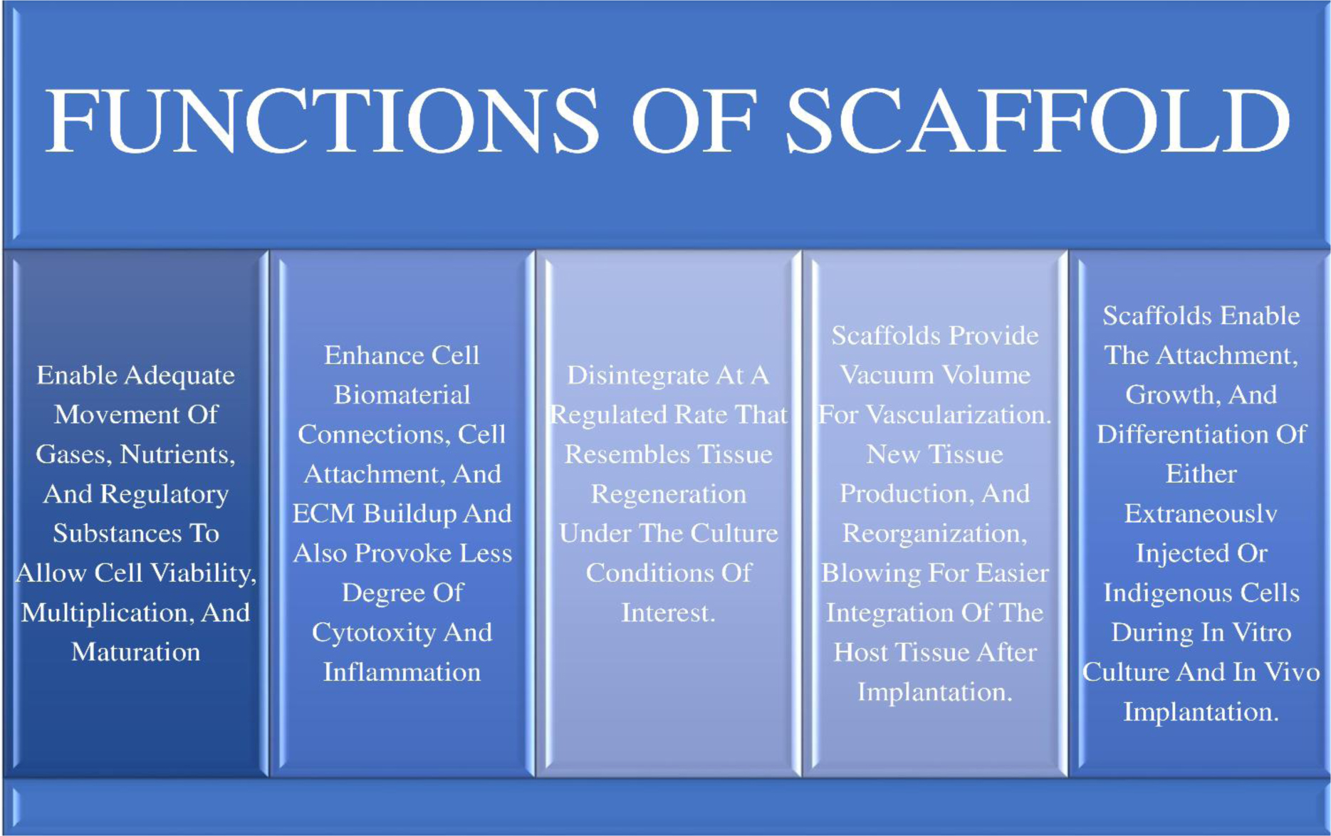
Craniofacial tissue-engineered techniques have significantly improved over the past 20 years as a result of developments in engineering and in material science. The regeneration of the craniofacial tissue is frequently complicated due to the craniofacial region's complexity, which includes bone, cartilage, soft tissue, and neurovascular bundles. It is now possible to construct tissues in the lab using scaffolds, cells, and physiologically active chemicals. For bone repair/augmentation, the biomaterials are classified into natural like “collagen, fibrin, alginate, silk, hyaluronate, chitosan” and synthetic like “polyethyleneglycol, poly-e-caprolactone, polyglycolic acid” and some bioceramics “tricalcium phosphate, hydroxyapatite, biphasic calcium phosphate, and the bioactive glasses” along with metals certain (Titanium and Zirconia ) and as this is part of advanced tissue engineering in dentistry there are some bioactive restorative materials like mineral trioxide aggregate and biodentine. The newer advanced techniques like 3D printed templates present a framework for achieving the three pillars of tissue engineering: healing, rebuilding and rejuvenation. The field of tissue engineering has recently become interested in 3D printing, also known as “Additive Manufacturing”, which is a ground-breaking technique that allows for the printing of patient-specific scaffolds, medical devices, multiscale, biomimetic/intricate cytoarchitecture/function-structure hierarchies and multicellular tissues in complex microenvironments. Biopolymers use is dependent on meeting the criteria for various scaffolds, including mechanical integrity, thermal stability, chemical composition, along with biological properties. Researchers have developed a revolutionary 4D bioprinting technique using cell traction forces and they are used to develop intricate dynamic structures, smart medical devices, or complex human organs.
Citation: Tanishka Taori, Anjali Borle, Shefali Maheshwari, Amit Reche. An insight into the biomaterials used in craniofacial tissue engineering inclusive of regenerative dentistry[J]. AIMS Bioengineering, 2023, 10(2): 153-174. doi: 10.3934/bioeng.2023011
Craniofacial tissue-engineered techniques have significantly improved over the past 20 years as a result of developments in engineering and in material science. The regeneration of the craniofacial tissue is frequently complicated due to the craniofacial region's complexity, which includes bone, cartilage, soft tissue, and neurovascular bundles. It is now possible to construct tissues in the lab using scaffolds, cells, and physiologically active chemicals. For bone repair/augmentation, the biomaterials are classified into natural like “collagen, fibrin, alginate, silk, hyaluronate, chitosan” and synthetic like “polyethyleneglycol, poly-e-caprolactone, polyglycolic acid” and some bioceramics “tricalcium phosphate, hydroxyapatite, biphasic calcium phosphate, and the bioactive glasses” along with metals certain (Titanium and Zirconia ) and as this is part of advanced tissue engineering in dentistry there are some bioactive restorative materials like mineral trioxide aggregate and biodentine. The newer advanced techniques like 3D printed templates present a framework for achieving the three pillars of tissue engineering: healing, rebuilding and rejuvenation. The field of tissue engineering has recently become interested in 3D printing, also known as “Additive Manufacturing”, which is a ground-breaking technique that allows for the printing of patient-specific scaffolds, medical devices, multiscale, biomimetic/intricate cytoarchitecture/function-structure hierarchies and multicellular tissues in complex microenvironments. Biopolymers use is dependent on meeting the criteria for various scaffolds, including mechanical integrity, thermal stability, chemical composition, along with biological properties. Researchers have developed a revolutionary 4D bioprinting technique using cell traction forces and they are used to develop intricate dynamic structures, smart medical devices, or complex human organs.
Three-Dimensional printing
computer-aided design
additive manufacturing
three-dimensional printing
solid freeform fabrication
tissue engineering
collagen, chitosan and tricalcium phosphate
Poly Lactic-co Glycolic acid
glycosaminoglycan
Calcium Phosphate
Hydroxyapatite
Mesenchymal Stem Cell
Chitosan
Tricalcium Phosphate
Bone Morphogenetic protein
Silk Fibroin
phosphate-buffered saline
hexafluoroisopropano
natural fibers reinforced composites
Cellulose nanocrystal
Nano fibrillated Cellulose
Bacterial nanocellulose
Polyethyleneglycol
Poly-E-Caprolactone
Polyglycolic Acid
Bicalcium Phosphate
Titanium
Aluminium
Vanadium
Polyetherketoneketone
High entrophy alloys
Laser cladding
Laser-aided additive manufacturing
Laser-cladded high-entropy alloy coatings
Bioactive glass
Silicon Dioxide
Sodium Dioxide
Calcium Oxide
Phosphorus Pentaoxide
polyetheretherketone
polyaryletherketone
Bone tissue regeneration
Regenerative Medicine
Shape Memory Polymers
Shape Memory Alloys
Polyvinyl Alcohol

| [1] |
KTevlin R, McArdle A, Atashroo D, et al. (2014) Biomaterials for craniofacial bone engineering. J Dent Res 93: 1187-1195. https://doi.org/10.1177/0022034514547271 
|
| [2] |
Chan BP, Leong KW (2008) Scaffolding in tissue engineering: general approaches and tissue-specific considerations. Eur Spine J 17: 467-479. https://doi.org/10.1007/s00586-008-0745-3
|
| [3] |
Arif ZU, Khalid MY, Noroozi R, et al. (2022) Recent advances in 3D-printed polylactide and polycaprolactone-based biomaterials for tissue engineering applications. Int J Biol Macromo 218: 930-968. https://doi.org/10.1016/j.ijbiomac.2022.07.140
|
| [4] |
Hsu EL, Ghodasra JH, Ashtekar A, et al. (2013) A comparative evaluation of factors influencing osteoinductivity among scaffolds designed for bone regeneration. Tissue Eng Part A 19: 1764-1772. https://doi.org/10.1089/ten.tea.2012.0711
|
| [5] |
Keane TJ, Badylak SF (2014) Biomaterials for tissue engineering applications. Semin Pediatr Surg 23: 112-118. https://doi.org/10.1053/j.sempedsurg.2014.06.010
|
| [6] |
Jafari M, Paknejad Z, Rad MR, et al. (2017) Polymeric scaffolds in tissue engineering: a literature review. J Biomed Mater Res Part B: Appl Biomater 105: 431-459. https://doi.org/10.1002/jbm.b.33547
|
| [7] |
Schulte M, Schultheiss M, Hartwig E, et al. (2000) Vertebral body replacement with a bioglass-polyurethane composite in spine metastases–clinical, radiological and biomechanical results. Eur Spine J 9: 437-444. https://doi.org/10.1007/s005860000162
|
| [8] |
Elisseeff J, Puleo C, Yang F, et al. (2005) Advances in skeletal tissue engineering with hydrogels. Orthod Craniofac Res 8: 150-161. https://doi.org/10.1111/j.1601-6343.2005.00335.x
|
| [9] |
Arvidson K, Abdallah BM, Applegate LA, et al. (2011) Bone regeneration and stem cells. J Cell Mol Med 15: 718-746. https://doi.org/10.1111/j.1582-4934.2010. 01224.x
|
| [10] |
Moshiri A, Oryan A (2012) Role of tissue engineering in tendon reconstructive surgery and regenerative medicine: current concepts, approaches and concerns. Hard Tissue 1: 11. https://doi.org/10.13172/2050-2303-1-2-291
|
| [11] |
Khalid MY, Arif ZU (2022) Novel biopolymer-based sustainable composites for food packaging applications: a narrative review. Food Packag Shelf Life 33: 100892. https://doi.org/10.1016/j.fpsl.2022.100892
|
| [12] |
Khalid MY, Al Rashid A, Arif ZU, et al. (2021) Natural fiber reinforced composites: Sustainable materials for emerging applications. Results Eng 11: 100263. https://doi.org/10.1016/j.rineng.2021.100263
|
| [13] |
Arif ZU, Khalid MY, Sheikh MF, et al. (2022) Biopolymeric sustainable materials and their emerging applications. J Environ Chem Eng 10: 108159. https://doi.org/10.1016/j.jece.2022.108159
|
| [14] |
Arif ZU, Khalid MY, Noroozi R, et al. (2023) Additive manufacturing of sustainable biomaterials for biomedical applications. Asian J Pharm Sci 2023: 100812. https://doi.org/10.1016/j.ajps.2023.100812
|
| [15] |
Kang BJ, Kim Y, Lee SH, et al. (2013) Collagen I gel promotes homogenous osteogenic differentiation of adipose tissue-derived mesenchymal stem cells in serum-derived albumin scaffold. J Biomater Sci Polym Ed 24: 1233-1243. https://doi.org/10.1080/09205063.2012.745717
|
| [16] |
Ferreira AM, Gentile P, Chiono V, et al. (2012) Collagen for bone tissue engineering. Acta Biomater 8: 3191-3200. https://doi.org/10.1016/j.actbio.2012.06.014
|
| [17] |
Kadler K (2004) Matrix loading: assembly of extracellular matrix collagen fibrils during embryogenesis. Birth Defects Res C 72: 1-11. https://doi.org/10.1002/bdrc.20002
|
| [18] |
Quinlan E, Thompson EM, Matsiko A, et al. (2015) Functionalization of a collagen-hydroxyapatite scaffold with osteostatin to facilitate enhanced bone regeneration. Adv Healthcare Mater 4: 2649-2656. https://doi.org/10.1002/adhm.201500439
|
| [19] |
Zhou C, Ye X, Fan Y, et al. (2014) Biomimetic fabrication of a three-level hierarchical calcium phosphate/collagen/hydroxyapatite scaffold for bone tissue engineering. Biofabrication 6: 035013. https://doi.org/10.1088/1758-5082/6/3/035013
|
| [20] |
Noori A, Ashrafi SJ, Vaez-Ghaemi R, et al. (2017) A review of fibrin and fibrin composites for bone tissue engineering. Int J Nanomed 12: 4937. https://doi.org/10.2147/IJN.S124671
|
| [21] | Siddiqui N, Pramanik K (2015) Development of fibrin conjugated chitosan/nano β-TCP composite scaffolds with improved cell supportive property for bone tissue regeneration. J Appl Polym Sci 132: 41534. https://doi.org/10.1002/app.41534 |
| [22] |
Kim BS, Sung HM, You HK, et al. (2014) Effects of fibrinogen concentration on fibrin glue and bone powder scaffolds in bone regeneration. J Biosci Bioeng 118: 469-475. https://doi.org/10.1016/j.jbiosc.2014.03.014
|
| [23] | Wong M (2004) Alginates in tissue engineering. Biopolymer Methods in Tissue Engineering. Methods in Molecular Biology™ . Switzerland: Humana Press 77-86. https://doi.org/10.1385/1-59259-428-X:77 |
| [24] |
Venkatesan J, Bhatnagar I, Kim SK (2014) Chitosan-alginate biocomposite containing fucoidan for bone tissue engineering. Marine Drugs 12: 300-316. https://doi.org/10.3390/md12010300
|
| [25] |
Valente JFA, Valente TAM, Alves P, et al. (2012) Alginate based scaffolds for bone tissue engineering. Mater Sci Engi C 32: 2596-2603. 10.1016/j.msec.2012.08.001
|
| [26] |
Kim M, Jung WK, Kim G (2013) Bio-composites composed of a solid free-form fabricated polycaprolactone and alginate-releasing bone morphogenic protein and bone formation peptide for bone tissue regeneration. Bioprocess Biosyst Eng 36: 1725-1734. https://doi.org/10.1007/s00449-013-0947-x
|
| [27] |
Pina S, Rebelo R, Correlo VM, et al. (2018) Bioceramics for osteochondral tissue engineering and regeneration. Osteochondral Tissue Engineering. Advances in Experimental Medicine and Biology . Cham: Springer 53-75. https://doi.org/10.1007/978-3-319-76711-6_3
|
| [28] |
Li DW, He J, He FL, et al. (2018) Silk fibroin/chitosan thin film promotes osteogenic and adipogenic differentiation of rat bone marrow-derived mesenchymal stem cells. J Biomater Appl 32: 1164-1173. https://doi.org/10.1177/0885328218757767
|
| [29] |
Aliramaji S, Zamanian A, Mozafari M (2017) Super-paramagnetic responsive silk fibroin/chitosan/magnetite scaffolds with tunable pore structures for bone tissue engineering applications. Mater Sci Eng C 70: 736-744. https://doi.org/10.1016/j.msec.2016.09.039
|
| [30] |
Zhang W, Ahluwalia IP, Literman R, et al. (2011) Human dental pulp progenitor cell behavior on aqueous and hexafluoroisopropanol based silk scaffolds. J Biomed Mater Res A 97: 414-422. https://doi.org/10.1002/jbm.a.33062
|
| [31] |
Collins MN, Birkinshaw C (2013) Hyaluronic acid-based scaffolds for tissue engineering—a review. Carbohydr Polym 92: 1262-1279. https://doi.org/10.1016/j.carbpol.2012.10.028
|
| [32] |
Seol YJ, Lee JY, Park YJ, et al. (2004) Chitosan sponges as tissue engineering scaffolds for bone formation. Biotechnol Lett 26: 1037-1041. https://doi.org/10.1023/B:BILE.0000032962.79531.fd
|
| [33] |
Balagangadharan K, Dhivya S, Selvamurugan N (2017) Chitosan based nanofibers in bone tissue engineering. Int J Biol Macromol 104: 1372-1382. https://doi.org/10.1016/j.ijbiomac.2016.12.046
|
| [34] |
Shalumon KT, Sowmya S, Sathish D, et al. (2013) Effect of incorporation of nanoscale bioactive glass and hydroxyapatite in PCL/chitosan nanofibers for bone and periodontal tissue engineering. J Biomed Nanotechnol 9: 430-440. https://doi.org/10.1166/jbn.2013.1559
|
| [35] |
Wang F, Zhang YC, Zhou H, et al. (2014) Evaluation of in vitro and in vivo osteogenic differentiation of nano-hydroxyapatite/chitosan/poly (lactide-co-glycolide) scaffolds with human umbilical cord mesenchymal stem cells. J Biomed Mater Res A 102: 760-768. https://doi.org/10.1002/jbm.a.34747
|
| [36] |
Khalid MY, Al Rashid A, Arif ZU, et al. (2021) Recent advances in nanocellulose-based different biomaterials: types, properties, and emerging applications. J Mater Res Technol 14: 2601-2623. https://doi.org/10.1016/j.jmrt.2021.07.128
|
| [37] |
Arif ZU, Khalid MY, Sheikh MF, et al. (2022) Biopolymeric sustainable materials and their emerging applications. J Environ Chem Eng 10: 108159. https://doi.org/10.1016/j.jece.2022.108159
|
| [38] |
Burdick JA, Anseth KS (2002) Photoencapsulation of osteoblasts in injectable RGD-modified PEG hydrogels for bone tissue engineering. Biomaterials 23: 4315-4323. https://doi.org/10.1016/s0142-9612(02)00176-x
|
| [39] |
Lin WJ, Flanagan DR, Linhardt RJ (1999) A novel fabrication of poly (ϵ-caprolactone) microspheres from blends of poly (ϵ-caprolactone) and poly (ethylene glycol) s. Polymer 40: 1731-1735. https://doi.org/10.1016/S0032-3861(98)00378-4
|
| [40] |
Nair LS, Laurencin CT (2007) Biodegradable polymers as biomaterials. Prog Polym Sci 32: 762-798. https://doi.org/10.1016/j.progpolymsci.2007.05.017
|
| [41] |
Ramesh N, Moratti SC, Dias GJ (2018) Hydroxyapatite–polymer biocomposites for bone regeneration: A review of current trends. J Biomed Mater Res Part B 106: 2046-2057. https://doi.org/10.1002/jbm.b.33950
|
| [42] |
Yang C, Unursaikhan O, Lee JS, et al. (2014) Osteoconductivity and biodegradation of synthetic bone substitutes with different tricalcium phosphate contents in rabbits. J Biomed Mater Res Part B 102: 80-88. https://doi.org/10.1002/jbm.b.32984
|
| [43] |
Özcan M, Hämmerle C (2012) Titanium as a reconstruction and implant material in dentistry: advantages and pitfalls. Materials 5: 1528-1545. https://doi.org/10.3390/ma5091528
|
| [44] | Anil S, Anand PS, Alghamdi H, et al. (2011) Dental implant surface enhancement and osseointegration. Implant Dentistry—A Rapidly Evolving Practice . Europe: InTech 83-108. https://doi.org/10.5772/16475 |
| [45] |
Wen CE, Yamada Y, Shimojima K, et al. (2002) Novel titanium foam for bone tissue engineering. J Mater Res 17: 2633-2639. https://doi.org/10.1557/JMR.2002.0382
|
| [46] |
Özkurt Z, Kazazoğlu E (2011) Zirconia dental implants: a literature review. J Oral Implantol 37: 367-376. https://doi.org/10.1563/AAID-JOI-D-09-00079
|
| [47] |
Abhay SS, Ganapathy D, Veeraiyan DN, et al. (2021) Wear resistance, color stability and displacement resistance of milled PEEK crowns compared to zirconia crowns under stimulated chewing and high-performance aging. Polymers 13: 3761. https://doi.org/10.3390/polym13213761
|
| [48] |
Arif ZU, Khalid MY, ur Rehman E (2022) Laser-aided additive manufacturing of high entropy alloys: processes, properties, and emerging applications. J Manuf Process 78: 131-171. https://doi.org/10.1016/j.jmapro.2022.04.014
|
| [49] |
Arif ZU, Khalid MY, Al Rashid A, et al. (2022) Laser deposition of high-entropy alloys: a comprehensive review. Opt Laser Technol 145: 107447. https://doi.org/10.1016/j.optlastec.2021.107447
|
| [50] |
Arif ZU, Khalid MY, ur Rehman E, et al. (2021) A review on laser cladding of high-entropy alloys, their recent trends and potential applications. J Manuf Process 68: 225-273. https://doi.org/10.1016/j.jmapro.2021.06.041
|
| [51] |
Skallevold HE, Rokaya D, Khurshid Z, et al. (2019) Bioactive glass applications in dentistry. Int J Mol Sci 20: 5960. https://doi.org/10.3390/ijms20235960
|
| [52] |
Alqurashi H, Khurshid Z, Syed AUY, et al. (2021) Polyetherketoneketone (PEKK): An emerging biomaterial for oral implants and dental prostheses. J Adv Res 28: 87-95. https://doi.org/10.1016/j.jare.2020.09.004
|
| [53] |
Cheng X, Wan Q, Pei X (2018) Graphene family materials in bone tissue regeneration: perspectives and challenges. Nanoscale Res Lett 13: 289. https://doi.org/10.1186/s11671-018-2694-z
|
| [54] |
Borrelli MR, Hu MS, Longaker MT, et al. (2020) Tissue engineering and regenerative medicine in craniofacial reconstruction and facial aesthetics. J Craniofac Surg 31: 15-27. https://doi.org/10.1097/SCS.0000000000005840
|
| [55] |
Khalid MY, Arif ZU, Noroozi R, et al. (2022) 4D printing of shape memory polymer composites: A review on fabrication techniques, applications, and future perspectives. J Manuf Process 81: 759-797. https://doi.org/10.1016/j.jmapro.2022.07.035
|
| [56] | Thrivikraman G, Athirasala A, Twohig C, et al. (2017) Biomaterials for craniofacial bone regeneration. Dent Clin 61: 835-856. https://doi.org/10.1016/j.cden.2017.06.003 |
| [57] |
Khalid MY, Arif ZU, Ahmed W (2022) 4D printing: technological and manufacturing renaissance. Macromol Mater Eng 307: 2200003. https://doi.org/10.1002/mame.202200003
|
| [58] |
Arif ZU, Khalid M Y, Zolfagharian A, et al. 4D bioprinting of smart polymers for biomedical applications: Recent progress, challenges, and future perspectives. React Funct Polym 179: 105374. https://doi.org/10.1016/j.reactfunctpolym.2022.105374
|
| [59] |
Mijiritsky E, Ferroni L, Gardin C, et al. (2017) Porcine bone scaffolds adsorb growth factors secreted by MSCs and improve bone tissue repair. Materials 10: 1054. https://doi.org/10.3390/ma10091054
|
| [60] |
Shakya AK, Kandalam U (2017) Three-dimensional macroporous materials for tissue engineering of craniofacial bone. Brit J Oral Max Surg 55: 875-891. https://doi.org/10.1016/j.bjoms.2017.09.007
|


Figures(8) / Tables(1)

Tanishka Taori, Anjali Borle, Shefali Maheshwari, Amit Reche. An insight into the biomaterials used in craniofacial tissue engineering inclusive of regenerative dentistry[J]. AIMS Bioengineering, 2023, 10(2): 153-174. doi: 10.3934/bioeng.2023011


 DownLoad: